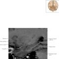
Thalamus and Basal Ganglia Axial 1

Normal Anatomy
The centrum semiovale, corona radiata, and internal capsules are all continuous white matter tracts. The centrum semiovale is the white matter deep to the gray matter on the surface of the brain and has an ovular shape. On axial imaging, it is generally a term used for white matter superior to the ventricles. The corona radiata (Latin: “sunburst”) is the white matter connecting the centrum semiovale superiorly and the internal capsules inferiorly. On axial imaging the corona radiata can be seen with ventricles. The internal capsule connects the corona radiata superiorly with the pyramids of the medulla inferiorly. On axial imaging, the internal capsule is between the caudate and lentiform nucleus anteriorly and the thalamus and lentiform nucleus posteriorly.

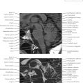
Thalamus and Basal Ganglia Axial 2

Normal Anatomy
The lentiform nucleus, a triangular-shaped structure on axial imaging, consists of the globus pallidus medially and the putamen laterally. The lentiform nucleus with the caudate nucleus constitutes the basal ganglia. The basal ganglia represent a large collection of nuclei that constantly modifies movement along with the cerebellum. The motor cortex sends information to both the cerebellum and basal ganglia, and both structures send information back to cortex via the thalamus (i.e., to gain access to the cortex, signals must pass through the thalamus).


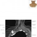
Thalamus and Basal Ganglia Axial 4