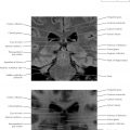
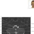
Limbic System Axial 2

Normal Anatomy
Note how the fornix (Latin, “arch” or “vault”) is more easily seen as a paired structure on the T2-weighted axial magnetic resonance image (upper radiology image), compared with the fornix as seen in the Chapter 3 images focusing on the basal ganglia and thalami. The fornix is a C -shaped bundle of axons in the brain and carries signals from the hippocampus to the hypothalamus.


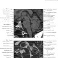
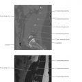
Limbic System Coronal 1