46.1 Multiple sclerosis: sagittal T2
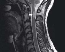
A focus of high signal intensity (arrow) is seen in the spinal cord. Clinically, this patient was suspected to have multiple sclerosis. which was confirmed by further lesions found in the brain
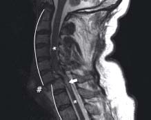
There is discontinuity (#) of the normal alignment due to C-spine fractures at C6 and C7. The spinal canal is markedly narrowed and there is high signal of the cord (arrow) which is a sign of oedema caused by cord injury. Normal cord is seen above and below (*)
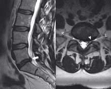
The sagittal image shows a low signal, degenerative and herniated L5/S1 intervertebral disc (short arrow). The axial view shows an intraforaminal protrusion (arrowhead) which is narrowing the left L5 exit foramen (L). Compare with the right (R)
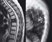
This patient with known breast cancer presented with back pain. There is a single metastasis in a mid-thoracic vertebral body. As the deposit is sclerotic (bony) it demonstrates low signal on all MRI sequences. For comparison the plain XR image shows increased density (arrowheads)
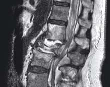
High signal is shown in two intervertebral discs due to infection (discitis) with destruction of the vertebral body between them. There is narrowing of the spinal canal at the level of the cauda equina leading to bilateral perianal and sciatic symptoms in this patient
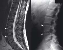
These are the ‘shiny corner’ or ‘Romanus’ lesions of early ankylosing spondylitis (arrowheads). The high signal (bright) represents oedema of the underlying marrow whereas the plain XR bright corners represent sclerosis of bone as it heals
Related posts:


Stay updated, free articles. Join our Telegram channel

Full access? Get Clinical Tree


