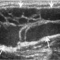
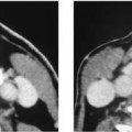
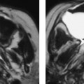
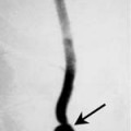
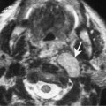
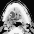
Chapter 62 Squamous cell carcinoma (SCCA) of the oral tongue is the second most common site of SCCA of the oral cavity. SCCA comprises > 95% of all malignancies of the oral tongue. The disease is more common in men between the ages of 50 and 70. The most common risk factors are smoking and excessive alcohol abuse. The staging of oral cavity carcinoma are presented in Table 62–1. The most common location is along the lateral border of the middle third of the oral tongue (Fig. 62–1). The common presenting symptoms include the sensation of a mass, a visible or palpable mass, local pain, dysphagia, or a palpable neck mass. Advanced tumors may extend posteriorly to invade the tongue base or inferiorly along the hyoglossus muscle and involve the floor of the mouth. This spread is often clinically occult. Bone erosion is unusual. Group I to II lymph nodes are at greatest risk for metastases. About 40% of patients will have palpable nodal disease and 20% will have bilateral involvement. Oral tongue lesions are usually ulcerative and infiltrative. SCCA of the oral tongue are usually moderately or well differentiated. They may arise in apparently normal epithelium; however, it is common to see these tumors associated with leukoplakia or chronic glossitis. The treatment for SCCA depends on the extent of disease. Wide local excision is performed for early lesions located along the lateral aspect of the oral tongue. Tumors that extend inferiorly into the floor of the mouth may require a marginal mandibulectomy. Tumors that cross the midline or extend posteriorly into the tongue base require a total or near-total glossectomy. The latter would also require a supraglottic laryngectomy to prevent the aspiration following the glossectomy. Because of the substantial morbidity associated with this procedure, combined chemotherapy and radiation therapy is gaining acceptance for advanced tumors that cross the midline or invade the tongue base.
Squamous Cell Carcinoma of the Oral Tongue
Epidemiology
Clinical Findings
Pathology
Treatment
TX | Primary tumor cannot be assessed |
T0 | No evidence of primary tumor |
