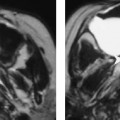
Chapter 70 The pharyngeal wall consists of the posterior and lateral pharyngeal mucosa that extends to the nasopharynx, superiorly, esophageal inlet, inferiorly, and tonsils laterally. The incidence is more common in males with the peak incidence between 60 and 70 years of age. The most common risk factors are excessive tobacco and alcohol use. Stages of oropharyngeal and hypopharyngeal cancer are presented in Tables 70–1 and 70–2, respectively. The most common complaint is sore throat. As these tumors progress (Fig. 70–1), patients may complain of dysphagia, foreign sensation, ear pain, blood-tinged saliva, aspiration, and change in voice quality. Patients with advanced disease may present with weight loss or a palpable neck mass indicative of a nodal metastasis.
Squamous Cell Carcinoma of the Posterior Pharyngeal Wall
Epidemiology
Clinical Findings
TIS | Carcinoma in situ |
T1 | Tumor ≤ 2 cm in greatest diameter |
T2 | Tumor > 2 cm but ≤ 4 cm in greatest diameter |
T3 | Tumor > 4 cm in greatest diameter |
T4a | Tumor invades larynx, deep/extrinsic muscle of tongue, medial pterygoid, hard palate, or mandible |
T4b | Tumor invades lateral pterygoid muscle, pterygoid plates, lateral nasopharynx, or skull base or encases carotid artery |