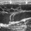
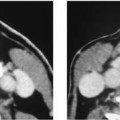
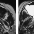
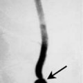
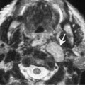
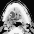
Chapter 68 The pyriform sinus is a subsite of the hypopharynx and is the most common location for squamous cell carcinoma (SCCA) that originates in the hypopharynx. The superior margin of the pyriform sinus is the pharyngoepiglottic fold, which is at the level of the base of the vallecula. The pyriform sinus tapers to an apex, which is located at the top of the cricoid cartilage. On cross-sectional imaging, the apex of the pyriform sinus is located at the level of the cricoarytenoid joint. The incidence of SCCA of the pyriform sinus cancer is lower than that of SCCA of the larynx. Risk factors include excessive tobacco and alcohol use, prior radiation therapy, and Plummer-Vinson syndrome (Paterson–Brown Kelly syndrome). This syndrome is thought to be precancerous and is characterized by iron-deficiency anemia; achlorhydria; generalized atrophy of the mucous membranes of the mouth, pharynx, and esophagus; webs in the hypopharynx and cervical esophagus; and weight loss. SCCA most commonly arises in middle-aged males. Plummer-Vinson syndrome is more common in women and should be considered in women with pyriform sinus or other types of hypopharyngeal carcinoma. The staging of hypopharyngeal carcinoma is presented in Table 68–1. The hypopharynx is often a clinically silent area for early tumors. As a result, many of these tumors are advanced at initial presentation. Symptoms associated with early tumors include persistent sore throat, or pain after swallowing hot foods or liquids. Advanced lesions often present with dysphagia, change in voice, enlarging neck mass, and weight loss. Referred ear pain (otalgia) may occur and is due to invasion of the superior laryngeal nerve (Fig. 68–1). It is important to note that up to 15% of patients will have a synchronous or metachronous second tumor.
Squamous Cell Carcinoma of the Pyriform Sinus
Epidemiology
Clinical Findings
Primary tumor cannot be assessed | |
T0 | No evidence of primary tumor |
Tis | Carcinoma in situ |
T1 | Tumor limited to one subsite of hypopharynx and < 2 cm in greatest dimension |
T2 | Tumor invades more than one subsite of hypopharynx or an adjacent site, or 2–4 cm in greatest diameter without fixation of hemilarynx |
T3 | Tumor > 4 cm in greatest diameter with fixation of hemilarynx |
T4a | Tumor invades thyroid/cricoid cartilage, hyoid bone, thyroid gland, esophagus, or central compartment soft tissue (prelaryngeal strap muscles and subcutaneous fat) |
T4b |
|
