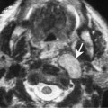
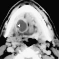
Chapter 59 The retromolar trigone (RMT) is a triangular area situated behind the molars between the posterior portion of the maxillary and mandibular alveolar ridges. Overall, the RMT is the least common site of squamous cell carcinoma (SCCA) of the oral cavity. However, it is one of the most frequently encountered oral cavity tumors associated with chewing betel nut. RMT SCCA is more common in men between the ages of 40 and 60. In our experience, RMT SCCA is one of the most difficult lesions to accurately stage by clinical examination. The stages of SCCA have been classified by AJCC (Table 59–1). A tumor that is felt to be early stage disease is often only “the tip of the iceberg.” These tumors commonly spread deeply to the pterygomandibular raphe and may continue to spread laterally along the buccinator muscle (Fig. 59–1). The RMT is located in close proximity to the anterior tonsillar pillar (palatoglossus muscle). As a result, these tumors may invade the palatoglossus muscle and extend superiorly to the soft palate or inferiorly to the tongue base. Early bone erosion is common and is often present without trismus. A common complaint is local and referred pain to the external auditory canal. Trismus is usually caused by extension into the masticator space. RMT SCCA may demonstrate perineural spread. The most common pathways are antegrade along the inferior alveolar nerve into the mandible, or retrograde along the inferior alveolar nerve to the main trunk of V3. The ipsilateral Group I lymph nodes are the primary echelon drainage for RMT SCCA. The incidence of clinically positive lymph nodes at presentation is between 35 and 40%.
Squamous Cell Carcinoma of the Retromolar Trigone
Epidemiology
Clinical Findings
TX | Primary tumor cannot be assessed |
T0 | No evidence of primary tumor |
Tis | Carcinoma in situ |
T1 | Tumor < 2 cm in greatest dimension |
