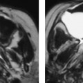
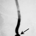
Chapter 67 The subglottic larynx extends from the inferior border of the true vocal cords to the inferior margin of the cricoid cartilage. Another commonly used definition to identify the proximal margin of the subglottis is that it begins mucosally at about 5 mm below the level of the free margin of the true vocal cords. Primary squamous cell carcinomas (SCCAs) of the subglottis are rare and constitute between 4 and 6% of all laryngeal carcinomas. Because of this rarity, it is difficult to determine the specific population at risk. However, it is probably similar to the patient population at risk for other types of SCCA of the larynx. The staging of subglottic carcinoma is presented in Table 67–1. Stridor and dyspnea are the most common symptoms. Hoarseness is also a common presenting complaint because many primary subglottic carcinomas are advanced at initial presentation and will involve the true vocal cords. Advanced lesions often extend inferiorly into the proximal trachea. Invasion of the cricoid cartilage and anterior extension through the cricothyroid membrane into the soft tissues of the neck are common (Fig. 67–1). The primary echelon lymph nodes for the subglottic larynx differ from other subsites of the larynx. The nodes at greatest risk of metastases from a primary SCCA of the subglottic larynx are the prelaryngeal (Delphian), pretracheal, paratracheal, and supraclavicular lymph nodes. The risk of nodal metastases is approximately 10%. Histologically, SCCA is classified as well, moderately, and poorly differentiated. On microscopic examination, SCCA appears as anaplastic cells found below the basement membrane with a variable degree of keratin production and intracellular bridges. Besides SCCA, other squamous cell aberrations that may arise in the larynx include the following categories: benign hyperplasia, benign keratosis, atypical hyperplasia, keratosis with epithelial atypia, intraepithelial carcinoma, and microinvasive squamous cell carcinoma. These lesions are unable to be distinguished by cross-sectional imaging. SCCA of the subglottis tend to be circumferential lesions that may extend superiorly to involve the true vocal cords and inferiorly to involve the proximal trachea. Advanced tumors may extend into the anterior soft tissues of the neck. Invasion of the cricoid cartilage is common.
Squamous Cell Carcinoma of the Subglottis
Epidemiology
Clinical Findings
Pathology
Related posts:


Stay updated, free articles. Join our Telegram channel

Full access? Get Clinical Tree


