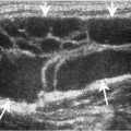
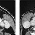
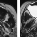
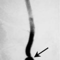
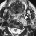
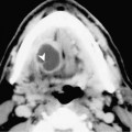
Chapter 57 The tongue base is located posterior to the oral tongue. The circumvallate papilla separates the oral tongue anteriorly (oral cavity) from the tongue base (oropharynx) posteriorly. It is important to differentiate the tongue base from the oral tongue because the anatomic subsite, lymphatic drainage, treatment, and prognosis are distinctly different. The posterior portion of the tongue base becomes continuous with the vallecula. Therefore, the squamous cell carcinomas (SCCAs) of the tongue or valleculae are often considered together and classified as a subsite of the oropharynx. SCCA comprises over 95% of all malignancies of the tongue. The disease is more common in men between the ages of 50 and 70. The most common risk factors are smoking and excessive alcohol abuse. The stages of SCCA have been classified by the American Joint Committee on Cancer (Table 57–1). The most common presenting symptoms include a visible or palpable mass, local pain, dysphagia, or a palpable neck mass. These tumors are often clinically silent and are often quite advanced at presentation. SCCAs of the tongue base are usually exophytic ulcerative tumors that tend to infiltrate beneath the mucosa. Early lesions are usually lateralized; however, advanced lesions often cross the midline. These tumors may also extend to the anterior tonsillar pillar, pharyngeal wall, submucosally under the valleculae into the supraglottic larynx, or anteriorly into the sublingual space. Lesions may also grow inferiorly and laterally to spread into the deep soft tissues of the neck, eventually involving the styloid musculature and the internal carotid artery (Fig. 57–1). As with all oropharyngeal carcinomas, bone erosion is unusual. The lymphatic drainage of the tongue base consists of a superficial and deep muscular lymphatic network. The superficial network is continuous with the superficial plexus that covers the oral tongue. The primary drainage is to Groups II and III. The deep lymphatic drainage may drain ipsilaterally or have direct branches that drain to the contralateral neck. There is a high likelihood for nodal metastases from tongue base carcinoma. Approximately 70% of patients will have either unilateral or bilateral cervical metastases at initial presentation.
Squamous Cell Carcinoma of the Tongue Base/Valleculae
Epidemiology
Clinical Findings
Tis | Carcinoma in situ |
T1 | Tumor ≤ 2 cm in greatest diameter |
T2 |
