7 Superficial Lymph Node Biopsy
Beth S. Edeiken-Monroe, Brett J. Monroe, Bruno D. Fornage, and Suhas G. Parulekar
- Ultrasound and ultrasound-guided needle biopsy of the regional nodal basins are important components of the initial staging of cancer patients. They
- Provide prognostic information
- Help in the formulation of the treatment plan
- Have proved accurate in diagnosing regional nodal involvement particularly in the groin, axilla, and neck
- Their major contribution and impact on management is the detection and diagnosis of early clinically occult ipsilateral or contralateral regional nodal metastasis that otherwise would not be included in the surgical dissection or radiation treatment field.
Indications for ultrasound guided biopsy of a lymph node include:
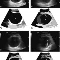
- Absence or displacement of the hilum
- Focal or diffuse thickening of the cortex
- Relative decreased echogenicity of the cortex
- Full or rounded shape of the lymph node
- Calcification within the lymph node
- Abnormal extrahilar vascular flow
- A cystic component
- Size is not a valid indication of an abnormal lymph node with the exception of asymmetric enlarged nodes in patients with lymphoma.
Ultrasound-guided fine-needle aspiration biopsy (FNAB) and core needle biopsy (CNB) are performed in the same room as ultrasound examinations in the outpatient facility. Ultrasound-guided FNAB is quick, relatively inexpensive, minimally invasive, and accurate when performed in the presence of an experienced cytopathologist.
- Focal metastatic deposits, as small as a few millimeters, can be targeted.
- In mixed solid/cystic lymph nodes, solid areas should be targeted.
- In patients with primary papillary thyroid cancer, fluid aspirate from a cystic lymph node may require determination of the thyroglobulin content of the fluid aspirate to diagnose metastatic disease.
- May be technically difficult to perform on small lymph nodes that are close to vessels or locations difficult to reach
- More traumatic than FNAB and is associated with an increased risk of hematoma formation
- Rarely used to diagnose metastatic adenopathy
- May be needed to provide pretreatment information in patients with breast cancer such as histologic grade, expressions of estrogen and progesterone receptors and c-erbB2
- In patients with suspected lymphoma, CNB may be necessary to diagnose small cell lymphoma.
- There are no absolute contraindications to the performance of an ultrasound-guided needle biopsy of a superficial lymph node.
- Anticoagulation – Risk of hemorrhage is reduced by halting anticoagulation therapy prior to biopsy for the recommended amount of time for each specific anticoagulant.
At our institution, the preprocedure evaluation includes:
- Allergy history
- Reconciliation of medications and vital signs with particular focus on blood pressure and anticoagulant medication
Ultrasound-guided needle biopsy is performed with:
- Standard ultrasound equipment
- Standard hypodermic needles
- Syringe for FNA
- An automated or semiautomated CNB device for CNB
- Ultrasound examination is performed using a high resolution ultrasound scanner with color and power Doppler capability equipped with commercially available high-frequency (from 7 MHz) linear–array transducers.
- FNAB is performed with standard hypodermic needles varying from 20–25 gauge attached to a 10 or 20 mL syringe depending on the preference of the operator.
- We routinely perform FNAB with a 20-gauge needle attached to a 20 mL syringe. This allows sufficient aspiration on a minimum of passes, usually only one.
- Alternatively, an 18-gauge needle is employed in the case of fibrotic node or node with a thick necrotic content.
Various sizes of core biopsy needles can be used ranging from 14-gauge to 20-gauge with the length of the throw ranging from 1.1–2.3 cm and the core sample length from 0.7–1.7 cm; the selection of the needle often depends on the location and size of the targeted node.