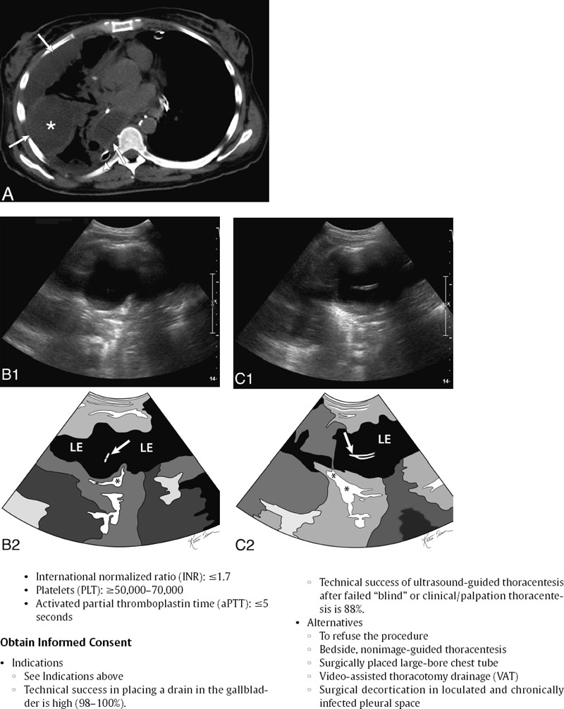
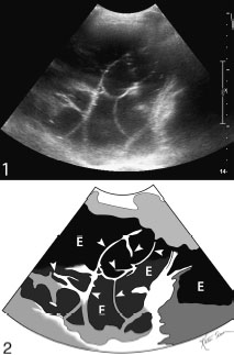
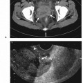
Thoracentesis

17 Thoracentesis


Wael E.A. Saad

Indications


By Presentation


  • Shortness of breath
  • Question of infection (fever)
  • Asymptomatic effusion of unknown etiology (diagnostic)
  • Recurrent effusions requiring pleurodesis (percutaneous access to pleural space)

By Etiology/Fluid Type


  • Pleural effusion

    • Transudate

      • Congestive heart failure
      • Renal failure
      • Hepatic hydrothorax
      • Hypoproteinemia
      • Meig’s syndrome

    • Exudate

      • Malignant effusion
      • Infections (including tuberculosis)

  • Pleural space infection

    • Empyema (from pneumonia, lung abscess)
    • Postoperative infection
    • Posttranshepatic procedures (bile leak, for example)

  • Hemothorax

    • Spontaneous
    • Malignant
    • Trauma
    • Postoperative

  • Chylothorax

    • Posttraumatic
    • Iatrogenic

Contraindications


Relative Contraindication


  • Uncorrected coagulopathy (relative)
  • Small amount of fluid for therapeutic procedure (for therapeutic intentions)
  • Mechanical ventilation is not a contraindication.

Preprocedural Evaluation


Evaluate Prior Cross-Sectional Imaging


  • Evaluate

  • Prior ultrasound images/evaluations of the pleural space may show septations within the pleural fluid (Fig. 17.2).


    image


    Fig. 17.2 Gray-scale ultrasound image of a septated pleural effusion (top) and schematic sketch of it (bottom). Numerous septa are seen (arrowheads) crisscrossing the pleural effusion (E).


    • The septations may indicate that a simple tube/drain placement may not adequately drain all the fluid in the pleural space.
    • This is an indication that the septations may require wire break-up during the procedure (see Fluoroscopic-guided wire and tube access).

  • Look for adjacent organs that can be inadvertently traversed

    • Transgression of organs is not a common problem.
    • It may be more common with small amounts of sub-pulmonic fluid, which raise the technical difficulty of a thoracentesis.
    • Organs that may be transgressed include the lung, spleen (left thoracentesis), and liver (right thoracentesis).
    • Plan a thoracentesis that is safe and away from adjacent structures such as the descending thoracic aorta particularly in left-sided posterior thoracentesis in the elderly with ectatic aorta

Evaluate Preprocedure Laboratory Values


Obtain Informed Consent


Equipment


Ultrasound Guidance


  • Multiarray 4–5 MHz ultrasound transducer
  • Transducer guide bracket (usually not necessary)
  • Sterile transducer cover

Standard Surgical Preparation and Draping


  • Chlorhexidine skin preparation/cleansing fluid
  • Fenestrated drape

Local Infiltrative Analgesia Administration


  • 21-gauge infiltration needle
  • 10– 20 mL 1% lidocaine syringe

Sharp Access Devices


  • 11-blade incision scalpel
  • 18-gauge needle that allows a 0.035-inch or 0.038-inch wire
  • 21-gauge needle that allows a 0.018-inch wire

Tubular Access Devices


  • Telescoped graduate dilation system (micropuncture kit transitional dilator) to upsize a 0.018-inch wire to a 0.035-inch wire
  • An 8-French fascial dilator that can be passed over a 0.035-inch guidewire may be used (see Fluoroscopic-guided wire and tube access).
  • An 8-French self-retaining (string-locking) pigtail drainage catheter, which is the definitive cholecystostomy tube/drain to be placed last (final product of the procedure)
  • All-in-one sharp and tubular access. This is a tube loaded coaxially on a hypodermic needle, which leads with a spring-loaded blunt tip. Once an indicator shows that there is fluid at the needle tip, the outer plastic Teflon tubing is passed over the needle and into the pleural space. For the purposes of this chapter, the Seldinger technique will be described.

Suction/Water Seal Devices


  • A thoracic tube in the pleural space can be drained utilizing sealed suction bottles (1 L vacuum bottles).
  • The thoracic drain/chest tube can be put to water seal or be on controlled suction (common level is -20-cm water wall suction) utilizing a pleurovac system; for example, chest drain multipurpose model Oasis (Atrium Medical Corp., Hudson, NH).

Technique

Stay updated, free articles. Join our Telegram channel

Mar 10, 2016 | Posted by in ULTRASONOGRAPHY | Comments Off on Thoracentesis

Full access? Get Clinical Tree

Get Clinical Tree app for offline access