Sutures and Fontanelles: Widened Sutures as a Symptom of Defective Ossification
10.1055/b-0034-87891
Sutures and Fontanelles: Widened Sutures as a Symptom of Defective Ossification
In this instance, no signs of increased ICP are present. Usually, the suture has sharp edges without prominent interdigitations. Suture widening is often associated with numerous wormian bones and a persistent fontanelle.
Rare causes of widened sutures include the following:
Pseudotumor cerebri, brain edema of unknown cause, post-traumatic with headache and papilledema: normal US, CT, and magnetic resonance imaging (MRI) findings
Superior sagittal sinus (SSS) thrombosis: diagnosed by US (Doppler), CT, or MRI
Vitamin A toxicity or deficiency
Hyperparathyroidism
Long-term use of steroid medication
Long-term prostaglandin E1 therapy
Lead poisoning.
Table 4.2 Widening of the sutures as a symptom of defective ossification
Diagnosis
Findings
Florid rickets
Indistinct suture margins, generally obscured osseous structures.
Hypothyroidism, untreated
Delayed closure of fontanelles, often numerous wormian bones.
Osteogenesis imperfecta
Numerous wormian bones are generally present; thinned calvarial bones.
Delayed closure of fontanelles. Syndrome is characterized by birdlike face, micropthalmia, cataracts, micrognathia, beaked nose, abnormal dentition, hypotrichosis, cutaneous atrophy, and proportional small stature.
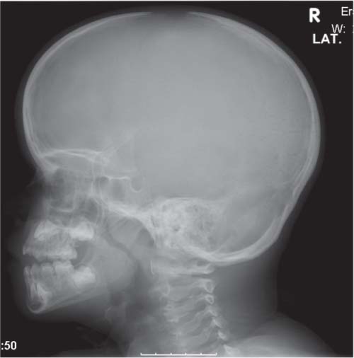
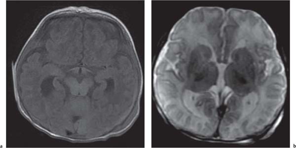
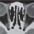
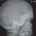
Fig. 4.2 Cleidocranial dysostosis in 4-year-old boy. Note the persistent open anterior fontanelle.Fig. 4.3a, b Epilepsy in a 2-day-old infant. Note the large open anterior fontanelle and facial manifestations that are suspected to be part of Zellweger syndrome.Fig. 4.4a, b Subependymal heterotopia as part of Zellweger syndrome on T1- and T2-weighted brain images of the same patient as Fig. 4.3.
Only gold members can continue reading. Log In or Register to continue