and Vincent L. Sorrell2
(1)
Division of Nuclear Medicine and Molecular Imaging Department of Radiology, University of Kentucky, Lexington, Kentucky, USA
(2)
Division of Cardiovascular Medicine Department of Internal Medicine Gill Heart Institute, University of Kentucky, Lexington, Kentucky, USA
Understanding the normal biodistribution and kinetics of the three most common cardiac radiopharmaceuticals (99mTc sestamibi, 99mTc tetrofosmin, 201Tl chloride) is fundamental to the practice of gamma camera-based MPI. The 99mTc radiopharmaceuticals localize into the myocardium more slowly with a 60 % first-pass extraction fraction compared to 85 % for 201Tl. They enter the myocardial cells by passive diffusion; uptake is proportional to blood flow at the time of the injection and cellular mitochondrial content. They are cleared by the liver, thus gallbladder and intestinal visualization is expected; the urinary tract is the secondary route of physiologic elimination. The hepatic clearance of 99mTc tetrofosmin is more rapid compared to 99mTc sestamibi. Conversely, as a potassium analog, 201Tl chloride is actively transported into the myocardial cells via the Na-K ATPase pump; it is cleared primarily by the urinary system and the gastrointestinal route is the alternate route. Another important difference is that 201Tl chloride redistributes relatively quickly (within minutes), while the 99mTc radiopharmaceuticals redistribute relatively slowly (4 h or longer) (Chamarthy and Travin 2010; Henzlova et al. 2009; Holly et al. 2010; Wackers et al. 1989).
The gamma camera instrumentation may be dedicated to imaging the heart with two small perpendicularly oriented detectors, or it may be a system with general purpose large field-of-view detectors that can be configured for SPECT MPI. The axis of rotation of the detectors is typically 180° from the right anterior oblique position to the left posterior oblique position in order to image the left-sided heart. Typically, for a 180° axis, 32 raw planar images are acquired by each of the two detectors (for a total of 64 images) with computer reconstruction of the projection data to create the SPECT MPI images. Some gamma camera systems are capable of attenuation correction, using either a radionuclide or an x-ray source, to correct for anterior soft-tissue attenuation (e.g., breast tissue) or inferior soft-tissue attenuation (e.g., diaphragm).
During image acquisition, the perfusion data may be gated to the R-R interval of the ECG monitor to create gated SPECT images. Gated SPECT images permit evaluation of global and regional left ventricular function and regional wall thickening. The end-systolic and end-diastolic left ventricular volumes and left ventricular ejection fraction (LVEF) can be calculated using automated contour software available from multiple vendors. The validity of these reported values are dependent upon the accuracy of the contours.
There are many accepted SPECT MPI stress test protocols (ACR–SNM–SPR 2009; Henzlova et al. 2009; Hesse et al. 2005; Holly et al. 2010; Strauss et al. 2008). Traditionally, patients undergo two scans, one during rest and the other after stress. Most protocols use the same intravenous radiopharmaceutical for both scans; however, some combine 201Tl chloride at rest with a 99mTc radiopharmaceutical for the stress test. Patients are stressed either by treadmill exercise or with one of several available pharmacologic agents (adenosine, regadenoson, dipyridamole, or dobutamine). Administered activities of the 99mTc MPI radiopharmaceuticals typically range from 7 to 12 mCi (259 to 444 MBq) at rest, with an approximate threefold increase from 22 to 30 mCi (814 to 1110 MBq) during stress for a one-day protocol. Morbidly obese patients undergoing two-day protocols are typically administered up to 40 mCi (1480 MBq) each day. For a 201Tl chloride stress/rest (redistribution) protocol, a single intravenous injection of 3–4 mCi (111–148 MBq) is required. Scan patterns, incidental findings, and imaging artifacts may vary considerably depending on the protocol followed. For example, the degree of gallbladder visualization is influenced by the fasting state which is usually different on a two-day stress/rest protocol compared to a one-day rest/stress protocol.
Regarding image acquisition, intravenous injection-to-imaging time intervals are quite variable. Traditionally, imaging commences immediately after stress for 201Tl chloride because of its rapid redistribution; delayed redistribution images are acquired 3–4 h later. After intravenous administration of a 99mTc radiopharmaceutical, the interval ranges anywhere from 15 to 90 minutes, with longer intervals after pharmacologic stress compared to exercise to allow for greater liver clearance (because increased splanchnic blood flow occurs during pharmacologic stress compared to exercise, thereby delivering more radiopharmaceutical to the liver). Persistent liver activity often results in cardiac reconstruction processing artifacts; thus, it is important to maximize liver clearance before cardiac imaging (Germano et al. 1994). Various strategies have been proposed to optimize liver clearance (Cherng et al. 2006).
Gastric activity is usually related to duodenogastric bile reflux and can be quite problematic because of the reconstruction processing artifacts related to the “hot” gastric fundus adjacent to the inferior myocardial wall. To remedy this confounding effect, different strategies to reduce or eliminate gastric activity have been tried with variable success (Boz et al. 2003; Hassan et al. 1991; Hofman et al. 2006; Hurwitz et al. 1993; Malhotra et al. 2010; Middleton and Williams 1994, 1996; Rehm et al. 1996; van Dongen and van Rijk 2000; Weinmann and Moretti 1999). No intervention is able to eliminate completely the possibility, and therefore, the reader must be able to recognize this finding and understand the potential ramifications.
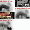
Processing of the raw projection data is generally performed with iterative reconstruction. Limits are set around the heart and the long-axis from base to apex is defined. Currently, dedicated computer software packages for SPECT MPI and gated SPECT analysis are standard and readily available from the vendors. In selected situations, reconstruction of the entire field-of-view data set—without limiting the reconstruction to the heart (“whole-field reconstruction” as is routine in noncardiac SPECT)—can be helpful to clarify and localize an extracardiac finding (Gedik et al. 2007; Seo et al. 2005). Figure 10.2 is one representative example highlighting the value of this additive technique.
Stay updated, free articles. Join our Telegram channel

Full access? Get Clinical Tree


