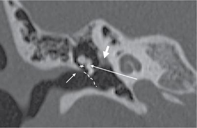
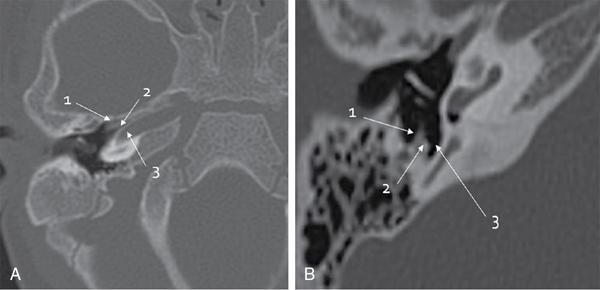
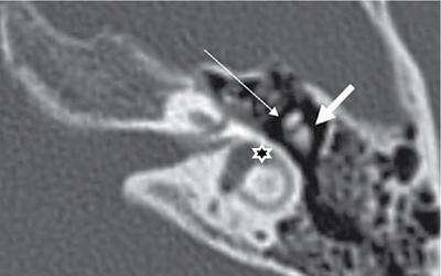
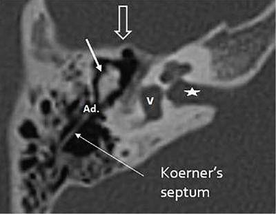
Rajendra Solanki, Pooja Prajapati, Poornima Digge, Digish Vaghela The ear mainly consists of three compartments: the inner, middle and external ear. However, embryologically it has dual development with inner ear develops from thickened ectodermal placode which receives neural sound. The external and middle ear develops from first and second pharyngeal arches and receives mechanical sound. It has very complex anatomy where multiple important structures are located in its close proximity like internal carotid artery, jugular bulb, sigmoid sinus, middle meningeal artery as well as seventh and eighth cranial nerves. Thus, understanding of its anatomy is very important. The external auditory canal is ‘S shaped’ structure which extends from pinna laterally to the tympanic membrane (TM) medially. It looks like a trumpet which flares out externally at pinna. Postero-superiorly, it is surrounded by mastoid portion of temporal bone, and antero-inferiorly, it is surrounded by tympanic portion of temporal bone. The external auditory canal has two portions: the outer one-third portion is fibrocartilaginous and inner 2/3rd portion is osseous. The two portions are connected by isthmus which is relatively narrow. Anteriorly, it contains the anterior tympanic recess which is located superior to eustachian tube. Scutum is a bony prominence located superiorly at the junction of external and middle ear. The lateral mallear ligament extends from scutum to the neck of malleus. The fibrocartilaginous portion bears defects in inferior aspect called ‘fissure of Santorini’, infections and metastases can pass through this deficiency. The medial portion sometimes have defect in anteroinferior area called ‘foramen of Huschke’ through which external auditory canal connects with temporomandibular joint. The TM is an oblique structure and forms boundary between middle and external ear (Fig. 3.30.1). It is conical in shape and measures 0.9–1 mm in diameter. It has two parts, pars tensa and pars flaccida. Pars tensa is rigid structure than flaccida due to a central fibrous layer. Pars flaccida is smaller than pars tensa and is superiorly located above the short process of malleus. The TM has a peripheral fibrocartilaginous ring which is deficient superiorly, above the short process of malleus, called notch of Rivinus. Prussak’s space: It is a small space located medial to pars flaccida and scutum, lateral to neck of malleus and superior to lateral malleal ligament fold. It is the most common site for the acquired cholesteatoma. The blunted scutum is hallmark of pars flaccida cholesteatoma. The middle ear is an air-filled cavity located between eustachian tube anteriorly and mastoid air cells posteriorly. Boundary of mesotympanum is as follows: The middle follows rule of three, that is., Three important sections, three ossicles, three structures in anterior wall and three structures in posterior wall (Fig. 3.30.2A and B). Two artificial imaginary planes divide middle ear cavity into three compartments: the hypotympanum, mesotympanum and epitympanum (Fig. 3.30.3). The hypotympanum and mesotympanum are divided by an artificial plane joining the tympanic annulus and cochlear promontory in the axial plane. The same way, mesotympanum and epitympanum are divided by an artificial plane extending between the scutum and tympanic segment of the facial nerve. The epitympanum contains aditus and antrum which leads to mastoid air cells posteriorly. Laterally, it has the lateral epitympanic space between the malleus and the scutum. Medially, there is a bony otic capsule which contains lateral semi-circular canal. The tegmen tympani forms the superior margin of the epitympanum and separates it from middle cranial fossa. Anteriorly, it communicates with epitympanic recess. It contains head of malleus, short process of incus as well as suspensory ligament. Axial section through epitympanum gives an ‘ice cream cone’ appearance, which corresponds to head of malleus and body of incus. Here, head of malleus articulates with body of incus and forms incudomalleal joint (Fig. 3.30.4). Located posterior to middle ear and external ear, it is subdivided into the mastoid antrum, which communicates with the epitympanum via the aditus, the central mastoid tract and the peripheral mastoid area. ‘Koerner septum’ is a thin bony structure which is formed by petrosquamous suture that extends posteriorly from the epitympanum, traversing through mastoid air cells and separating mastoid air cells into medial (petrous) and lateral (squamous) compartments (Fig. 3.30.5). It easily gets eroded in cholesteatoma. The mesotympanum is biggest compartment of tympanic cavity contains the main portion of the ossicular chain. The ossicular chain is composed of three bones: malleus (head, neck, anterior process, lateral process, and handle), incus (body, short process, long process, and lenticular process), and stapes (head/capitellum, anterior crus, posterior crus, and footplate). Apart from these, few ligaments and two muscle, tensor tympani and stapedius muscles also seen, which insert on the malleus and stapes, respectively. The posterior wall contains three important structures, the sinus tympani, facial recess and pyramidal eminence. The handle of the malleus is attached to the TM, the bone forms two joints, incudomalleal joint (between body of incus and head of malleus) which is located in epitympanic region and the incudostapedial joint (between long process of incus and head of stapes). Foot process of stapes attaches to oval window. The ossicles are best appreciated in axial plane. However, due to complexity and smaller structures, certain reconstruction techniques and landmarks helps for better recognition which are as follows.
3.30: Temporal bone imaging anatomy
Introduction
External auditory canal
Tympanic membrane
Middle ear
Anterior Wall (carotid wall)
Contains an opening of eustachian tube, semi canal for tensor tympani muscle and it separates middle ear from internal carotid artery.
Inferior wall (jugular wall)
Separates tympanic cavity from superior bulb of internal jugular vein.
Superior wall (tegmental wall)
Formed by tegmen tympani which separates middle ear from middle cranial fossa. It contains canal for tensor tympani muscle and mastoid antrum.
Lateral wall (membranous wall)
Formed by tympanic membrane
Posterior wall (mastoid wall)
Contains the canal of facial nerve, superiorly the bulge of semi-circular canal and pyramid, below to the aditus which is pierced by tendon of stapedius muscle.
Medial wall (labyrinthine wall)
Separates middle ear from inner ear. It contains 5 structures from superior to inferior are – Bulge of lateral semi-circular canal, facial canal bulge, Oval window, promontory (basal turn of cochlea) and round window.

Epitympanum
Mastoid region
Mesotympanum
Ossicular chain
Related posts:


Stay updated, free articles. Join our Telegram channel

Full access? Get Clinical Tree


