| CRANIAL REGION | Tentorium |
| HISTOPATHOLOGY | N/A; radiographic diagnosis of dural arteriovenous fistula |
| PRIOR SURGICAL RESECTION | No |
| PERTINENT LABORATORY FINDINGS | N/A |
Case description
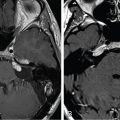
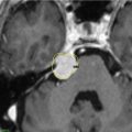
The patient is a 60-year-old male who presented with acute onset of aphasia and headache. Magnetic resonance imaging (MRI) was performed ( Figure 12.62.1 ), and diffusion-weighted images showed a right temporoparietal infarct with hemorrhagic conversion. He underwent digital subtraction angiography, which revealed a small tentorial dural arteriovenous fistula (DAVF) associated with cortical venous drainage (CVD). The patient subsequently underwent Gamma Knife radiosurgery (GKRS) with a 23-Gy marginal dose at the 50% isodose line ( Figure 12.62.2 ).
| Radiosurgery Machine | Gamma Knife – Perfexion |
| Radiosurgery Dose (Gy) | 23, at the 50% isodose line |
| Number of Fractions | 1 |

MRI showed a lesion suspicious of dural arteriovenous fistula (DAVF). This was confirmed on digital subtraction angiography, which revealed a small tentorial DAVF associated with cortical venous drainage (arrow).

GKRS plan of the DAVF with a prescribed marginal dose of 23 Gy at the 50% isodose line. Angiography was performed on the day of the procedure before the radiosurgery for accurate localization of the nodules. GKRS, Gamma Knife radiosurgery; DAVF, dural arteriovenous fistula.
| Critical Structure | Dose Tolerance |
|---|---|
| Transverse sinus | Not known |
Related posts:
 Esthesioneuroblastoma – delayed postoperative radiosurgery for recurrence at long-term
 Null cell – delayed postoperative radiosurgery for growing perioptic residual
 Chondrosarcoma – definitive radiosurgery after subtotal resections
 Large vestibular schwannoma – delayed postoperative radiosurgery for growing residual
 Trigeminal neuralgia due to petroclival meningioma – upfront radiosurgery
 Superior sagittal sinus meningioma – immediate postoperative radiosurgery for residual
Stay updated, free articles. Join our Telegram channel

Full access? Get Clinical Tree


