Chapter 87
Teratoma
Epidemiology
Teratomas are true neoplasms of germ cell origin that consist of elements derived from all three germ cell layers. The incidence is 1 in 4000 births. Teratomas may occur in numerous locations in the extracranial head and neck, including the neck, paranasal sinuses, nasopharynx, orbit, and pharynx. Seven to nine percent of teratomas occur in the cervical region. There is no reported gender predilection for cervical teratomas. However, nasopharyngeal teratomas have been reported to have a strong female predilection (6:1).
Clinical Findings
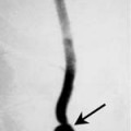
Cervical teratomas usually present in the first year of life and are often detected at birth. These lesions are often identified on prenatal ultrasound. They may be seen in full-term, premature, and stillborn infants. Cervical teratomas present as a bulky neck mass that may cross the midline. The symptoms of the mass are usually due to compression of adjacent structures. Tracheal compression may result in stridor, apnea, or cyanosis. Compression of the esophagus may result in dysphagia. About 20% of affected infants are associated with maternal polyhydramnios. This may be due to the inability of the infant to swallow amniotic fluid in utero due to esophageal compression.
Embryology
The embryogenesis of cervical teratomas is controversial. Historically, these lesions were felt to arise from anomalous development of the thyroid gland because they contained thyroid tissue. However, the explanation of the origin of cervical teratomas arising from the thyroglossal duct or the pharyngeal pouches (ultimobranchial anlage) is debatable.
Pathology
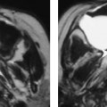
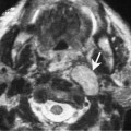
The majority of teratomas are benign lesions that contain histologically identifiable tissue from all three germ layers. Neural tissue is identified in 75 to 85% of cervical teratomas. These lesions typically have a well-defined capsule. The presence of fat and calcification allows easy diagnosis on imaging studies. Approximately one third of cases will have foci of normal thyroid tissue within the teratoma wall.