, Valery Kornienko2 and Igor Pronin2
(1)
N.N. Blockhin Russian Cancer Research Center, Moscow, Russia
(2)
N.N. Burdenko National Scientific and Practical Center for Neurosurgery, Moscow, Russia
Testicular cancer does not exceed 2% by the frequency of metastases in the brain (Guenot 1994). The clinical picture of metastatic lesions in PrC and testicular cancer is not different from the clinical picture of metastases of cancers from other sites in the brain. We observed two cases of brain involvement in this cancer (less than 1%).
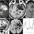
CT studies conducted in patients with cerebral metastases of testicular cancer did not identify any specific radiological characteristics and pathognomonic symptoms as compared to those in metastatic brain lesions of other primary tumors. In a series of similar cases, the authors noted that solitary metastases were found at the site of the primary tumor in the pelvic area (86%)—prostate and urinary bladder—in the half of cases. The metastatic lesions identified have the density identical to or lower than that of the brain; however, in case of an isodense structure, the presence of a metastatic lesion can be confirmed by the edema area surrounding the tumor. The contrast agent accumulation has a ring-shaped character with the adjacent tissue component—“ring + tissue.”
Stay updated, free articles. Join our Telegram channel

Full access? Get Clinical Tree


