The Adrenal Glands


The Adrenal Glands


Amber Wooten


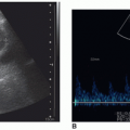
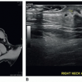
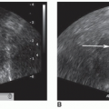
In contrast with other examinations of the abdomen, transabdominal sonographic imaging is not the first-choice imaging modality for screening adrenal glands or detecting adrenal pathology. Examination of the adrenal glands challenges the skills of novice and experienced sonographers alike and requires a working knowledge of exact adrenal anatomic locations and landmarks. However, combining planar flexibility and improved beam-resolution capabilities of modern ultrasound equipment with maneuvering the patient into multiple positions increases the likelihood of producing quality sonographic images of the adrenal glands.1, 2 and 3 In addition, since the mid-1990s, endoscopic ultrasound (EUS) and intraoperative ultrasound (IOUS) have developed as tools for high-resolution sonography evaluation of suprarenal and retroperitoneal masses. Using a 7.5-MHz transducer positioned 1 to 2 cm from the adrenal gland, EUS and IOUS are particularly useful for detecting adrenal metastases and staging cancer.4,5 As new technologies are introduced for transabdominal imaging, such as three-dimensional/four-dimensional (3D/4D) imaging and elastography, these tools also become available for EUS and IOUS applications.6,7

This chapter provides an overview of relevant anatomy, embryology, scanning techniques, functional and morphologic adrenal pathology, and the sonographic appearance
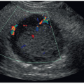
of the normal gland as well as adrenal pathology. As a sonographer, it is important to develop critical-thinking and problem-solving skills for sorting out the origin and extent of abdominal masses, including those of suprarenal and retroperitoneal origin. Related techniques are discussed. In addition, EUS, IOUS, computed tomography (CT), magnetic resonance imaging (MRI), and associated nuclear medicine imaging procedures are presented as related to adrenal imaging. Owing to technical improvements in all the diagnostic imaging modalities and the increased use of these modalities, more clinically silent or unexpected masses are being detected. The general term for an unexpected mass detected during an imaging procedure being performed for unrelated disease is “incidentaloma.” This term may be applied to any unexpected mass occurring anywhere in the body. Adrenal incidentaloma (AI) has become a common term in the medical literature and represents a diagnostic challenge that includes discovery of masses, which range from benign, nonfunctional lesions to malignant tumors such as pheochromocytomas, adrenocortical carcinomas, or metastases from other primary cancers.8 Sonographers have a role both in the initial detection of AIs and in gathering diagnostic information regarding newly discovered AIs.


ANATOMY


Embryology

The adrenal gland consists of two distinct parts: the cortex and the medulla. Each develops from different embryonic tissues, forms different anatomic and functional structures, and combines within a common capsule.9,10 The result is two endocrine glands in one organ. Most glands of the body develop from epithelial tissue. In contrast, the adrenal cortex is derived from the mesoderm of the same region that gives rise to gonadal tissue.9,10 The central tissue or adrenal medulla is functionally part of the sympathetic nervous system developed from the neural crest cells that also give rise to postganglionic sympathetic neurons.9, 10 and 11 As a result, some adrenal medullary pathology may appear ectopically along the paths of these sympathetic neurons usually near the celiac axis, whereas ectopic adrenocortical tissue may be located inferiorly along the path of gonadal tissue migration.9, 10 and 11 In addition, extra-adrenal chromaffin cells also normally deposit near the aortic bifurcation to form the organ of Zuckerkandl.10, 11 and 12


Cortex

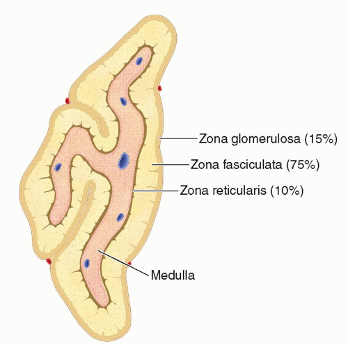
During gestational weeks 5 and 6, the fetal cortex is first recognized bilaterally as a groove between the developing dorsal mesentery and gonad.9,10 During weeks 7 and 8, the cells arrange into cords with dilated blood spaces, forming a thin capsule of connective tissue that encloses the gland9,10 and develops an intimate relationship with the superior pole of the kidney. If the kidney does not develop normally, there will be a discoid distortion in the shape of the adrenal gland.9,10 Initially, the fetal adrenal gland is larger than the kidney and 10 to 20 times the relative size of the adult adrenal gland.9,10 During the remainder of fetal life, the cortical tissue is composed of two zones comprising 75% to 80% of the bulk of the gland.10,11 By week 8, the cortex produces precursors to androgen, estriol, and corticosteroids.9,10 After birth, the inner zone undergoes involution, whereas the thinner outer zone continues to develop into the adult adrenal cortex, which takes on a yellow color.9, 10, 11, 12 and 13 By the age of 3 years, the cortex differentiates into three zones: (1) zona glomerulosa, (2) zona fasciculata, and (3) zona reticularis. Each zone develops different cellular arrangements and becomes functionally specialized, producing mineralocorticoids, glucocorticoids, and gonadocorticoids, respectively9, 10, 11 and 12,14 (Fig. 15-1).


Medulla

Specific ectodermal cells ascend from the neural crest, migrate from their origin, and differentiate into sympathetic neurons of the autonomic nervous system.9, 10 and 11 Some of these primitive autonomic ganglia differentiate even further into endocrine cells, designated chromaffin cells, and migrate to form a mass on the medial surface of the fetal adrenal cortex.9, 10 and 11 Soon, these chromaffins, or pheochrome cells, invade the developing cortex, establishing the primordium of the adrenal medulla.9,10 On cut section, the medulla has a red, brown, or gray color depending on the level of blood perfusion.15 As mentioned previously, chromaffin cells also form the organ of Zuckerkandl.10, 11 and 12


Relational Anatomy

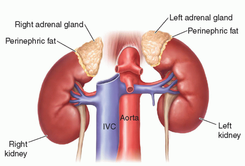
Like the kidneys, the adrenal glands are retroperitoneal. They are generally anterior, medial, and superior to the kidneys.9,10,13,15 The cortex and medulla are encapsulated by a thick inner layer of fatty connective tissue.12,13 A thin, fibrous outer capsule attaches to the gland by many fibrous bands, providing the adrenals with their own fascial supports so that they do not descend if the kidneys are displaced or absent.9,12 The glands are attached to the anteromedial aspect within renal fascia (also referred to as Gerota fascia), and abundant adipose tissue (perinephric fat) surrounds each gland, separating it from the kidneys11,12,16 (Fig. 15-2).


Right Adrenal

The right adrenal gland is located posterior and lateral to the inferior vena cava (IVC), medial to the right lobe of
the liver and lateral to the crus of the diaphragm.9,10,13,15,16 The right adrenal gland has been described as sitting like a triangular cap on the anterior, medial, and superior aspects of the superior pole of the right kidney.


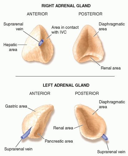
The anterior surface of the right adrenal gland is shaped like a pyramid. Two areas make up the anterior surface: the medial area is narrow and lies posterolateral to the IVC and the lateral, somewhat triangular, portion is in contact with the liver.9,10,13,15,16 The superior end of the lateral area is devoid of the peritoneum as it comes in contact with the bare area of the liver, and the inferior portion is covered by reflected peritoneum from the inferior layer of the coronary ligament12,13,15 (Fig. 15-3).

A curved ridge separates the posterior dorsal surface into superior and inferior parts. The superior convex portion rests on the diaphragm, and the inferior concave portion is in contact with the superior-anterior surface of the right kidney15 (Fig. 15-3).


Left Adrenal

The left adrenal gland appears draped in an elongated, crescent, or semilunar shape on the medial aspect of the left kidney’s superior pole.9, 10, 11, 12 and 13,15,16 The left gland is larger than the right, and extension to the left renal hilus is a normal variant.

The anterior portion can be separated into superior and inferior parts. The superior area is situated posterior to the peritoneal wall of the lesser sac and is covered by the peritoneum of the omental bursa, which separates the gland from the cardiac portion of the stomach.15 The inferior area is not covered by the peritoneum and lies posterior and lateral to the pancreas9,10,12,13,15,16 (Fig. 15-3). The splenic artery and vein course between the pancreas and the left adrenal gland.

The posterior surface is in close proximity to the splanchnic nerves.6,15 It is divided into a medial and a lateral area by a vertical ridge. The larger lateral area rests on the kidney and the medial posterior area on the crus of the diaphragm12,13,15 (Fig. 15-3).


Systemic and Lymphatic Vessels

Similar to other endocrine glands, the adrenals are among the most vascular organs of the body.13,15,16 The vasculature of the adrenal gland is distinguished from other organs in that the arteries and veins do not actually course together. The abundant arterial supply may contain as many as 50 to 60 small terminal arterioles, whereas the venous blood is channeled almost completely through a single, large venous trunk.12,13,15,16


Arteries

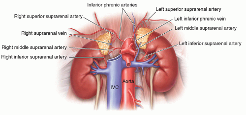
Three arteries supply each gland: the superiorly located suprarenal branch of the inferior phrenic artery, the superior and medially located branch of the aorta, and the inferiorly located suprarenal branch of the renal artery12,13,15,16 (Fig. 15-4).

These arteries are distinctively classified into three types: short capsular arterioles, intermediate cortical arteries (long branches that go through the cortex to the medulla), and the medullary sinusoids.15


Veins

In each adrenal gland, a central vein runs the length of the gland and exits at the hilum.12 The right suprarenal vein empties directly into the posterior aspect of the IVC as a short (4 to 5 mm) vessel, which exits the gland on the mid-anteromedial surface.12,15 The left suprarenal vein drains directly inferior and medial into the left renal vein.12,15 Frequently, the left inferior phrenic vein and the left suprarenal vein join before emptying into the left renal vein12,15 (Fig. 15-4).


Lymphatics

Lymph channels drain from the adrenal cortex and medulla to the hilar area.15 Following the arterial pathways, larger lymphatic vessels drain into para-aortic and lumbar lymph nodes, which drain to the cisterna chyli, thoracic duct, and eventually into the subclavian vein, whereas a few lymphatic vessels drain into the posterior mediastinal lymph nodes.12,17


PHYSIOLOGY

Just as their origin and structure are unique, the function and control of hormones differ for these two different glands in one organ. Adrenal pathology and some medications have the potential to disrupt the level of adrenal hormone secretion and associated regulatory mechanisms.11,14,18, 19 and 20 Hormones produced by the adrenal cortex, such as cortisol, are essential to life and must be replaced if both adrenal glands are removed.11,14,18, 19 and 20


Cortex

The cortex makes up 90% of the adrenal gland. By 3 years of age, the cortex develops into three epithelial layers, each one evolving functionally into very specialized zones, producing steroid hormones consistent with their mesodermal source. The zona glomerulosa, the outer layer directly beneath the connective tissue covering, makes up 15% of the cortex and produces aldosterone, a mineralocorticoid.9,10,13,14,16 The zona fasciculata, the middle layer, comprises 75% of the cortex and the zona reticularis, the inner layer, accounts for the remaining 10% of the cortex9, 10 and 11,13,16 (Fig. 15-1). Cortisol, a glucocorticoid, and two gonadocorticoids, estrogen and androgen, are produced by the zona fasciculata and zona reticularis.9,10,13,14,16

Hormone secretion is often controlled by the negative feedback mechanisms.14 Low blood concentrations of a hormone trigger the hypothalamus to secrete the primary regulating factor, corticotrophin-releasing hormone (CRH), which triggers the anterior lobe of the pituitary to release adrenocorticotropic hormone (ACTH).14 As blood concentrations of ACTH increase, adrenal hormone activity increases, producing a higher concentration of hormones, such as cortisol, in the bloodstream. This increased concentration of the adrenal hormone inhibits CRH and ACTH and, ultimately, hormone synthesis. When the blood concentration of one or more of the adrenal hormones drops to low levels, the cycle is repeated.14,18 Adrenocortical hormone secretion, function, and regulation are summarized in Table 15-1.


Medulla

Originating from ectodermal cells, the medulla secretes catecholamine hormones, similar to the posterior pituitary and thyroid glands. The medulla’s chromaffin (pheochrome) cells, the hormone-producing portion, surround large blood-filled sinuses.9,10,13,16

Epinephrine (adrenalin) and norepinephrine (noradrenalin) are the two principal hormones synthesized by the medulla. Epinephrine constitutes about 80% of the total secretion, and its action is more important than norepinephrine. Release of both hormones is usually stimulated through the sympathetic nervous system.11,14

Adrenal nerve stimulation results in prompt discharge of medullary hormones without materially influencing cortical secretion.11,14 Hormone secretion is controlled directly by the autonomic nervous system, and innervation by the preganglionic fibers allows the gland to respond to the neural stimulus.14 The anticipation or presence of stress or pain causes the hypothalamus to signal the sympathetic preganglionic neurons to stimulate the chromaffin cells to increase output of epinephrine and norepinephrine.11,14 Functionally, the medulla is a large sympathetic ganglion, which triggers action via hormone release instead of through axons.11,14 The body responds by (1) accelerating the heart rate and constricting the vessels, causing increased blood pressure; (2) accelerating the rate of respiration and dilating the respiratory passage; (3) decreasing the rate of digestion to make available more blood to the muscles, increasing the efficiency of muscle contraction; and (4) increasing the blood sugar level to provide energy, thus stimulating cellular metabolism.11,14 This physiologic response to stress is better known as the fight-or-flight response.11,14,18 Hypoglycemia, hypotension, hypoxia, hypovolemia, and exposure to temperature extremes may also stimulate medullary secretion of epinephrine and norepinephrine.11,14 Like the glucocorticoids of the adrenal cortices, these hormones help the body resist stress; however, unlike the cortical hormones, the medullary hormones are not essential to life.11,14


FUNCTION TESTS

There are many different kinds of laboratory tests to evaluate adrenocortical function. They can be divided into two types: tests that determine the absolute values in serum and urine versus tests that check the interdependency of the various hormones. Table 15-2 summarizes the significance of increased and decreased variance from normal laboratory values for various serum and urine tests.


Of the limited tests available for testing medullary function, 24-hour urine samples are typically used for catecholamines.11,14,18,20 Metanephrines are measured in urine, whereas dopamine may be assessed via urine or blood samples.18,20 With hypertension, pheochromocytoma, or neuroblastoma, urine levels of catecholamine, vanillylmandelic acid (VMA), or both may be elevated.11,14,18,20

Multiple tests are designed to determine the true functions and interdependency of the hypothalamus, pituitary, kidneys, and adrenals, including stimulation and suppression testing for ACTH, aldosterone, and cortisol.11,14,18,19


SONOGRAPHIC SCANNING TECHNIQUE


Preparation

Usually, patients receive no preparatory instructions for sonography of the adrenal glands, but when there is suspicion of a mass or metastases or a need to image any retroperitoneal anatomy sonographically, it is recommended that the patient fast approximately 6 to 8 hours before the examination.


Scanning Protocol

Evaluation of adrenals may be approached with the patient in supine or decubitus positions. Common protocols typically begin with transverse scanning, followed by longitudinal and/or coronal assessment. Slight or small scanning maneuvers are used to obtain desired images. One of the imaging goals for adrenal sonography is to document unilateral versus bilateral pathologic involvement. As with most sonographic examinations, width and anteroposterior measurements in transverse sections and length measurements from the longitudinal/coronal sections are required. Various breathing excursions and suspended inspiration may be used to optimize visualization of both adrenal glands.

The sonographer should select the highest-frequency transducer that will provide adequate penetration, given an adrenal depth range of 4 to 12 cm, depending on the patient’s size.21 Selection of a transducer with a small footprint will also facilitate scanning through intercostal spaces.1,3 Magnifying the field size may improve visualization of small structures. Scanning the patient in a prone position is uncommon, although this method is useful to show the spatial relationship between a large adrenal mass and the adjacent kidney.


Right Adrenal

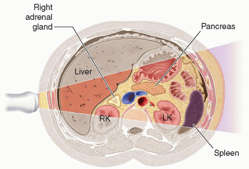
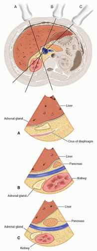
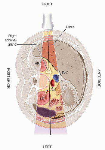
For imaging the right adrenal, the liver and sometimes the right kidney are useful acoustic windows (Fig. 15-5). Successful utilization of the liver as an acoustic window depends on hepatic size and attenuation characteristics (Fig. 15-6). Starting with transverse scans using an intercostal approach, the section of the IVC located medial and anterior to the upper pole of the kidney should be found.1,2 The plane should be directed toward the lateral and posterior aspects of this portion of the IVC while also keeping the ultrasound beam perpendicular to the spine. The adrenal gland should be seen in this location anterior to the crus of the diaphragm. The entire right adrenal gland is evaluated by scanning transversely from the renal hilus and proceeding superiorly (Fig. 15-5).


Longitudinal or coronal scanning of the right gland can be accomplished with several approaches1,3 (Fig. 15-6). A higher success rate has been reported when utilizing the liver as an acoustic window and scanning the patient in a left lateral decubitus position.6,22,23 From an intercostal window, the transducer is angled anterior toward the IVC and posterior toward the right kidney until the entire gland is visualized in the longitudinal or coronal plane (Fig. 15-7).


Left Adrenal

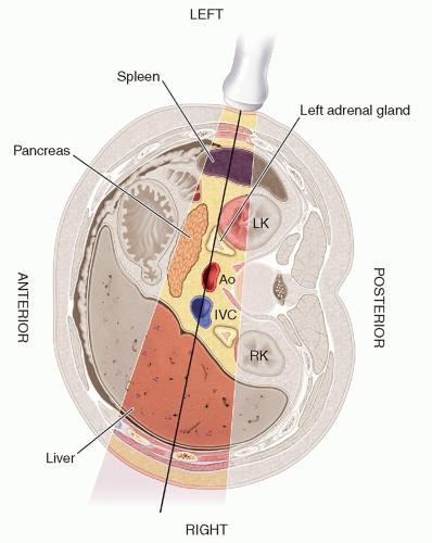
The left adrenal gland is more difficult to locate and document. Conventionally, the left adrenal has been imaged with the patient in the right decubitus position, using the spleen or left kidney as an acoustic window21,22,24 (Fig. 15-8). Identifying the left adrenal and its alignment is first done in the transverse plane. The left adrenal should lie between the left kidney and the aorta, with the pancreatic tail and splenic vein marking the superior margin of the gland.25

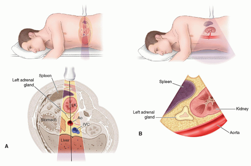
Longitudinal or coronal scans may be easier to obtain with the patient in a right anterior oblique position using a left posterior oblique scanning plane. First, in the transverse plane, the aorta medial and anterior to the upper pole of the left kidney or the spleen should be located, until the axis of the left kidney and the position of the aorta are determined and the left adrenal gland is identified between these structures22,23 (Fig. 15-9A). With the patient in the same position, the transducer should be rotated into the longitudinal/coronal plane. When imaging the left adrenal gland, the transducer may have to be oriented obliquely (Fig. 15-9B).

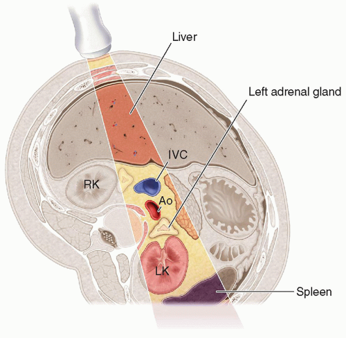
In the mid-1980s, Krebs and colleagues21,24 introduced an alternative approach to improve localization and delineation of the left adrenal gland. The patient is placed in a 45-degree left posterior oblique position, termed the cava-suprarenal line position.21,24,26 The transducer is placed on the patient’s right side, allowing the acoustic beam to pass through a double vascular acoustic window, the IVC, and the aorta21 (Fig. 15-10). The protocol starts with transverse scans until the left adrenal gland is located; longitudinal views follow. With this position, the success rate is 90%, compared with 60% in the same patient population using the conventional approach.21,24


Pitfalls

The size, location, and pathology of the adrenals and of surrounding structures impose significant limitations on sonographic visualization. Cirrhosis with fatty infiltration of the liver and obesity interfere with adequate penetration. Shadowing from the ribs and narrow intercostal spaces also make this approach challenging.

The right adrenal gland may be obscured by gas and food in the second portion of the duodenum. It is important to differentiate the crus of the diaphragm as a tubular structure located medial to the right adrenal, because it can be mistaken for a normal gland.27 The right adrenal gland is usually displaced posteriorly when the retroperitoneal fat line is displaced by liver disease.27,28

Structures that converge in the area of the left adrenal may mimic this gland: the esophagogastric junction, stomach, gastric diverticula, splenic vessels, portosystemic collateral vessels, tail of the pancreas, prominent hepatic lobes, medial lobulations of the spleen, superior lobulations of the kidney, or adjacent tumors.1,3,27,29, 30, 31 and 32 A more posterolateral approach or the cava-suprarenal line position may be indicated for proper visualization of the left adrenal gland.21,24


NORMAL SONOGRAPHIC ANATOMY

Fetal adrenal glands are quite large, and 90% of the time at least one can be imaged after 26 to 27 weeks of gestation.33 In adults, the adrenal glands are much smaller. The glands are generally located anterior, medial, and superior to the kidneys and vary in shape and configuration.11,32 Their size varies from 3 to 6 cm long, 2 to 4 cm wide, and 3 to 10 mm thick; the adult adrenals weigh 4 to 14 g.1,3,22,23,32,34 Transabdominally, the cortex and medulla are usually sonographically indistinguishable, because the normal internal texture appears homogeneous and hypoechoic.32 The glands are usually surrounded by highly echogenic fat, and in some patients, only the echogenic fat can be identified in contrast to an anechoic adrenal gland.32 Higher-resolution transducers such as those used for endoscopic and intraoperative applications are able to routinely detect the hyperechoic medullary echoes against the hypoechoic cortical echoes and the hyperechoic halo of fatty tissue.4,17


Right Adrenal

The right adrenal gland is identified superior to the kidney and lateral to the right crus of the diaphragm.23,32 On transverse sections, the gland is described as having a triangular, trapezoid, or inverted Y or V shape, with the tail extending from the anteromedial aspect of the right kidney3,30,32,35 (Fig. 15-11A). In a longitudinal plane scanning the medial aspect of the gland, the anteromedial ridge appears as a curvilinear or S-shaped structure and is visualized posterior to the IVC, slightly above or at the level of the portal vein.1,22,23,32,35 Moving laterally in longitudinal planes through the right adrenal gland, the anterior and posterior wings spread open and the gland takes on an inverted Y or V shape35 (Fig. 15-11B, C). The anterolateral portion is medial and posterior to the right lobe of the liver and posterior to the duodenum.32 Care should be taken to differentiate the right adrenal gland from the more medial hypoechoic/anechoic tubular right crus of the diaphragm.

Only gold members can continue reading. Log In or Register to continue

Stay updated, free articles. Join our Telegram channel

Dec 10, 2022 | Posted by in ULTRASONOGRAPHY | Comments Off on The Adrenal Glands

Full access? Get Clinical Tree
Get Clinical Tree app for offline access