Case presentation
A 12-year-old male presents with an injury to the chest. Apparently, he was “playing” with a nail gun and it went off, causing a nail to enter his chest. The child’s vital signs show an afebrile patient, who looks apprehensive, pale, and diaphoretic. His heart rate is 112 beats per minute, respiratory rate is 22 breaths per minute, blood pressure is 112/62 mm Hg, and pulse oximetry reading is 95% on room air. He has what appears to be a wound to his right chest, just lateral to the sternum at the nipple line, without active bleeding. His breath sounds are equal. He does not have any other injuries.
Imaging considerations
Pediatric penetrating chest trauma accounts for up to 8% of pediatric trauma cases. The majority of cases are seen in adolescent populations. Associated injuries include pneumothorax, hemothorax, cardiac, and great vessel injuries. When penetrating injury to the heart occurs, the right ventricle is more commonly involved and associated cardiac tamponade is often present. , Nail gun injuries in the pediatric population are uncommon. For a complete discussion on pediatric thoracic trauma, see Chapter 47 (Chest Pains: Pediatric Chest Trauma).
Plain radiography
Plain radiography is an excellent first-line imaging modality to identify the presence of radiopaque foreign bodies involved in penetrating thoracic injury and can identify clinically significant associated injuries, such as pneumothorax and hemothorax. In one case series of penetrating thoracic injury due to a nail gun, chest radiography was diagnostic in all patients; no further imaging was performed and the patients in this series all underwent urgent operative intervention.
Ultrasound
Echocardiogram can be used to identify pericardial effusions and assess ventricular function and can be useful in planning surgical intervention. , Retained foreign bodies can also be visualized and their location determined. Pericardial hemorrhage can be detected and alert the clinician to the presence of cardiac tamponade, which, in one review, was present in up to 73% of patients with cardiac nail gun injuries. The advantages of lack of ionizing radiation and availability make this imaging modality particularly attractive.
Computed tomography (CT)
CT may be useful in pediatric penetrating thoracic injury, as concern for blood vessel injury is higher in patients who sustain this traumatic mechanism. Consider utilization of this modality in children with suspected cardiovascular injuries. , It is also reasonable to consider employing chest CT with angiography in children with abnormal initial chest radiographs suggestive of aortic injury.
Imaging findings
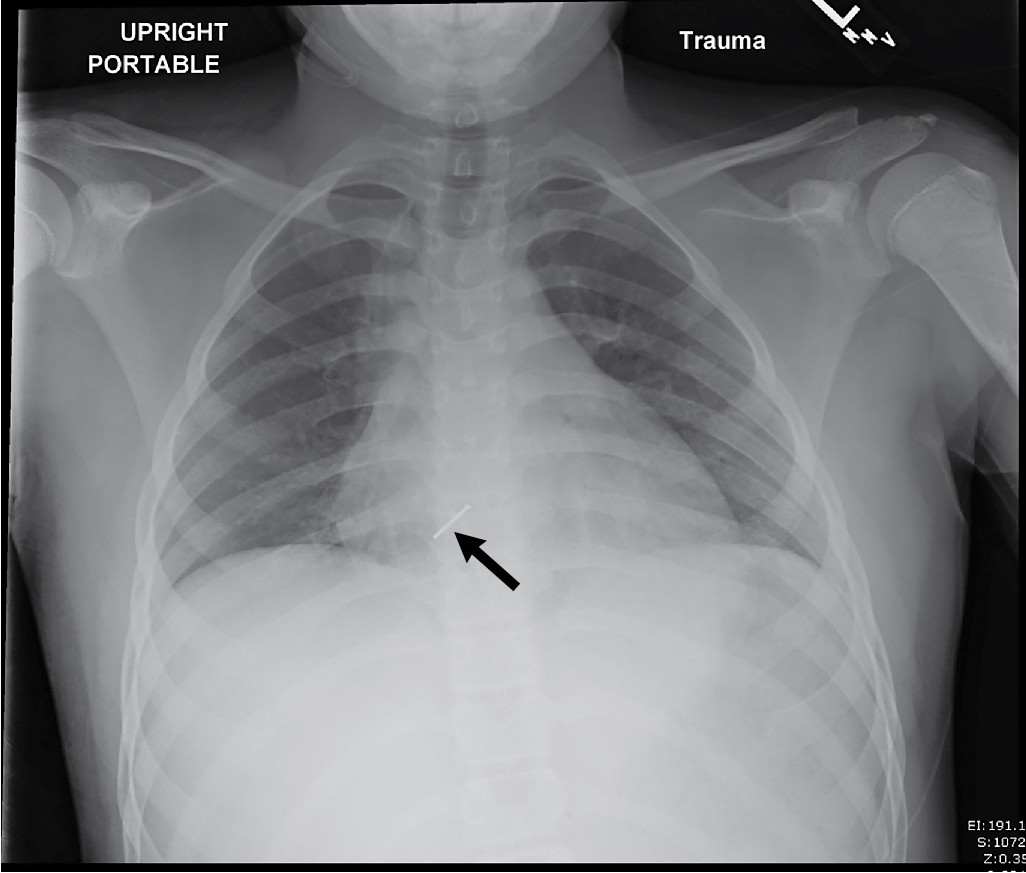
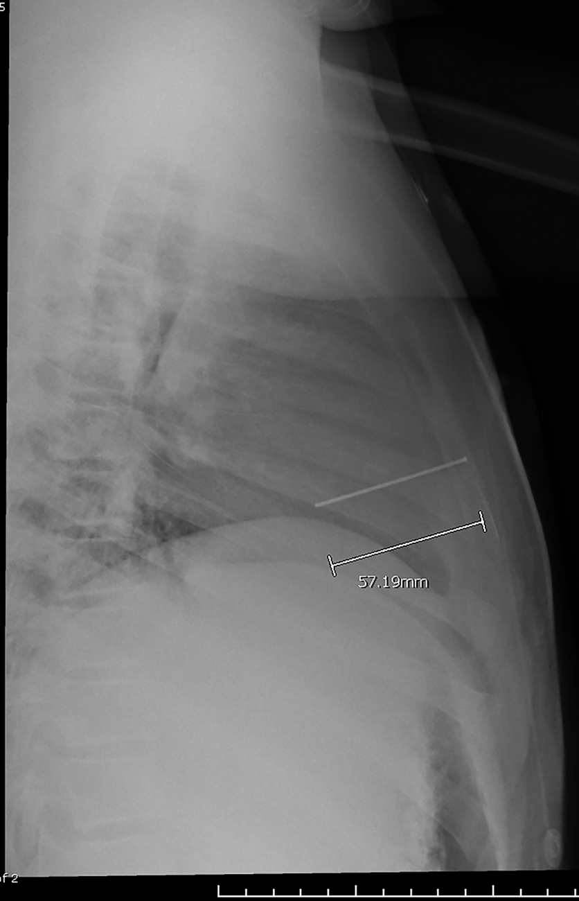
The child underwent immediate plain radiography of the chest. These images demonstrated a linear metallic object measuring 5.7 cm long overlying the right side of the cardiac silhouette, in the anterior chest. The location and orientation of the object suggest that it is intracardiac ( Figs. 46.1 and 46.2 ).