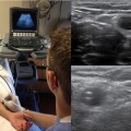
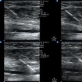
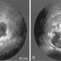
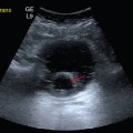
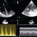
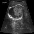
57 The approaches to examination of patients by clinical providers has evolved with the growth of medical knowledge and expectations of quality care. Though still primarily relying on their senses, physicians have been adding basic equipment (e.g., scale, stethoscope) and more complex devices (e.g., scopes, sphygmomanometer) to patient evaluation standards. In the intensive care unit (ICU), the diagnostic process is rather complex and continuous, and physical examination is deprived of several basic elements, such as pain assessment and general patient cooperation. Therefore, intensivists tend to rely on adjunctive diagnostic tools. The addition of critical care ultrasound (CCU) to the patient examination arsenal has been a major development during the last decade. The method fits naturally in the logic of the initial assessment process and, because of its repeatability, also in the continuous monitoring of these highly variable patients. As a generous source of real-time information, CCU shares a pedestal only with the physical examination itself.1,2 One overarching and powerful measure capable of tipping the balance toward accelerated maturation and phasing in of CCU is the development and promotion of a high-level, conceptually strong, and realistic approach. This chapter does that by further analyzing the holistic approach (HOLA) concept of CCU imaging that was introduced in Chapter 1. The HOLA concept defines CCU as part of the patient examination by a clinician to visualize all or any parts of the body, tissues, organs, and systems in their live, anatomically and functionally interconnected state and in the context of the whole patient’s clinical circumstances. The term “holistic” in the HOLA acronym is used in its original meaning in ancient Greek: to emphasize the importance of the whole and the interdependence of its parts. The term and the acronym must not be confused with “holistic medicine,” which has a different patient population, scope, and methodology. The authors of this chapter define the following overarching principles that should drive implementation of a HOLA-based CCU model in the ICU: 1. CCU is applicable in a “head-to-toe” fashion to follow and augment the process of physical examination and has an instantaneous effect on patient management. 2. Although specific CCU techniques deal with particular anatomic or pathologic entities, any tissue in any location in the body is subject to generic scanning. 3. A basic battery (profile) of CCU techniques can be adopted as a standard component of the bedside evaluation of every ICU patient. 4. Specialized batteries of CCU techniques can be implemented to best address the needs in specific clinical situations or patient categories. 5. Either CCU techniques or generic scanning should be categorized into basic and consultant levels. Basic techniques are performed by ICU team members. Consultant-level examinations that require radiology, cardiology, or other specialized expertise are supported by other teams external to the ICU but constitute a part of the overall imaging strategy in the ICU. 6. System- and facility-level acceptance is necessary for an appropriately planned and executed implementation process to gradually upgrade the conventional ICU to a “HOLA-capable” and, ultimately, an operational “HOLA-certified” status. 7. The ultimate stage of HOLA implementation is a full-fledged CCU laboratory with advanced equipment; clinical procedure support; archival, training, and broad quality assurance functions; and established interfaces with other hospital services and personnel. The scope of CCU is not to replace comprehensive sonography. CCU does not substitute for “radiologic” sonography, very much like Foley catheter placement does not make the urology service irrelevant; CCU replaces only studies that are largely futile in their solely retrospective significance. CCU performed by intensivists or other members of the ICU team augments the physical examination and has an instant effect on patient management. Moreover, it aids in dynamic monitoring of the constantly changing clinical status of ICU patients because of its bedside availability and repeatability. This dynamic monitoring capability is a distinct CCU feature that radiology routines can never adopt (e.g., recognize interstitial pulmonary edema and assess the effect of diuresis every 10 minutes). Most CCU techniques promise better outcomes only if used within the clinically driven time frame. Typically, still image–based, technician-performed studies are associated with delayed radiologist interpretation and serve different purposes. The HOLA concept of CCU imaging was introduced in Chapter 1. This textbook in its entirety is, in essence, a detailed description of HOLA. In all fairness, adoption of HOLA is a major, understandably difficult conversion of a physician’s mindset and attitude toward changing the established routine of clinical practice. It takes effort to reconsider the notion that scanning is a prerogative of equipment-savvy technicians and image interpretation is a separate “darkroom magic” by radiologists. Such conversion requires realization and acceptance of the unity and synchrony of scanning and interpretation when performed by a clinician to see or rule out pathologies in real-time by using somewhat unconventional techniques and implementing complex monitoring protocols.3
The holistic approach ultrasound concept and the role of the critical care ultrasound laboratory
Overview
Scope of critical care ultrasound
The HOLA concept of critical care ultrasound imaging
![]()
Stay updated, free articles. Join our Telegram channel

Full access? Get Clinical Tree


Radiology Key
Fastest Radiology Insight Engine

