The Neck


10.1055/b-0034-87904

The Neck


Table 4.47 Midline masses

Diagnosis


Findings


Comments


Thyroglossal duct syst


Fig. 4.158


US/CT/MRI: midline cystic mass embedded in infrahyoid strap muscles. Hypodense/intense and anechoic unless infected.


If calcified or has solid elements, needs work-up for malignancy.


Most present < 10 y.


US confirms normal thyroid gland.


CT/MRI if cyst suprahyoid or infected.


Ectopic thyroid


CT/MRI: Well-circumscribed midline or paramedian tongue mass with density/intensity similar to normal thyroid tissue. Can be multifocal.


May expand rapidly during puberty.


Assess normal thyroid tissue as lingual thyroid is only functioning tissue in 75% of cases.


Dermoid/epidermoid


CECT: cystic, well-demarcated mass containing fluid (epidermoid) or fat/fluid (dermoid).


MRI: Use fat-saturated techniques to diagnose dermoid.


Unusual before 5 y of age. Usually involve floor of mouth but if large, can protrude inferiorly.


Lymph node


US: typical node appearances.


(see Table 4.49 )


Cervical thymus


US/MRI: midline or on the left.


(see Table 4.49 )


Extension of plunging ranula


Fig. 4.159


US/CT: cystic.


MRI: Thin-walled cyst with intermediate signal on T1 and high signal on T2. Bulk of the lesion is in submandibular space with characteristic connection to the floor of the mouth.


Fig. 4.158 Transverse US of neck shows a hypoechoic midline cystic lesion consistent with a thyroglossal duct cyst.
Fig. 4.159 Axial postcontrast CT of the neck at the level of the angle of the mandible shows a right nonenhancing hypodense cystic lesion in the submandibular space consistent with a plunging ranula.


Table 4.48 Lateral cystic masses

Venolymphatic malformations


Fig. 4.160


Fig. 4.161


MRI: T2 for extent and fluid levels, postcontrast T1 with fat saturation for enhancing septations and venous elements.


CT: shows phleboliths.


Usually identified at or soon after birth. US helpful initially to direct further imaging.


1. Lymphatic malformation—transspatial cystic mass uni- or multilocular.


2. Venous malformation—enhancing mass with calcifications.


3. Lymphaticovenous—cystic and enhancing trans-spatial lesion.


Branchial cleft cyst


US: demonstrates location, usually hypoechoic.


CT/MRI: demonstrate infection with enhancing wall/increased attenuation and signal.


Cystic mass posterolateral to submandibular gland, lateral to carotid space, anterior or anteromedial to SCM muscle.


Abscess


Fig. 4.162, p. 394


US/CT/MRI: fluid-filled mass with irregular, thick enhancing wall and low-density/-intensity center. If involving thyroid, think of fourth branchial pouch anomaly.


Present with symptoms and signs of infection.


Thymic cyst


US/CT/MRI: Nonenhancing, low attenuation, hypo-/anechoic left lateral neck cyst closely associated with carotid sheath. Solid components rare, represent aberrant thymic/parathyroid tissue.


Can occur anywhere along thymopharyngeal duct but left lateral infrahyoid neck at level of thyroid gland is most common.


Laryngocoele


Radiograph: shows air-filled lateral structure. Best imaged with CT or MRI, which show unilocular air or fluid-filled cyst. Lies within the submandibular space.


Occur as a result of increased intraglottic pressure. Increase in size with Valsalva.


(Para) Thyroid cyst


Usually colloid cyst.


(see Table 4.53 )


Varix of jugular vein


US: expanded jugular vein with Doppler flow.


Best seen during straining, coughing, or crying.

Fig. 4.160 A large, left, multiseptated cystic mass displacing the carotid space posteriorly and airway medially is consistent with a venolymphatic malformation on axial postcontrast CT of the suprahyoid neck.
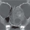
Fig. 4.161 A large hyperintense cystic mass without septations is consistent with a venolymphatic malformation on T2 coronal MRI of the face and neck.
Fig. 4.162 T1 axial postcontrast MRI of the suprahyoid neck shows left neck swelling with marked enhancement of soft tissues and a central rim-enhancing hypointense area consistent with an abscess.


Table 4.49 Lateral solid masses

Diagnosis


Findings


Comments


Fibromatosis colli


Fig. 4.163a, b


US: Focal mass in or diffuse enlargement of the middle/lower third of affected SCM muscle. Variable echogenicity.


CT: enlarged muscle without discrete mass.


MRI: Usually iso-/hypointense to normal muscle on T1. T2 variable but usually hypointense at area of maximal enlargement probably due to fibrosis.


Infant with torticollis tilting head to same side and rotating head toward opposite side.


Lymph nodes/lymphoma


Fig. 4.164


US: Oval nodes increased in size and number. Central hypoechogenicity in suppuration.


CECT: best to differentiate cellulitis/phlegmon/abscess and assess extent of disease in suspected malignancy.


MRI: T1 low/intermediate signal; T2 high signal.


Findings nonspecific and depend on clinical scenario; often rely on tissue diagnosis.


Teratoma


Fig. 4.130, p. 378


CT/MRI: Large and infiltrative with multiple cystic and solid elements. Contain fat and calcification. Both modalities show extent and components.


Usually < 1 y; may cause significant airway and feeding compromise.


Neuroblastoma


CT/MRI: Well-defined mass lying posterior to the vascular sheath and occasionally extending into the cervical spinal canal. MRI is best for delineating extent of tumor and spinal and intracranial extent.


Less than 5% occur in the neck.


Need chest and abdomen CT as part of initial work-up to rule out abdominal primary.


Hemangioma


US: well-defined mass with prominent vessels.


MRI: Best for extent. T1 isointense to muscle with intense enhancement postcontrast; T2 hyperintense with multiple flow voids.


Characteristic presentation; therefore, imaging indicated if suspect deep extension, pretreatment, or to assess treatment response.


Rhabdomyosarcoma


CT: heterogeneous lesion with or without osseous destruction.


MRI: isointense to muscle on T1, hyperintense on T2 and show enhancement postcontrast.


May be a primary lesion or related to metastatic adenopathy that is present in 50% of patients.


Forty percent occur in head and neck.


Fibrosarcoma/sarcoma


CT/MRI: Heterogeneously enhancing soft-tissue mass. Usually less intense and homogeneous enhancement than hemangioma. May show osseous destruction.


Often with regional lymph node involvement. Need chest CT for metastatic surveillance.


Hematoma


US: Imaging findings depend on age of bleed. Acute bleed hyper-echoic, less echogenic in 96 h, and eventually anechoic. Usually well defined but may become irregular if blood escapes into adjacent structures. Differentiate from tumor by absence of flow.


Usually due to trauma, anticoagulation, or coagulopathy.


Neurofibroma/plexiform neurofibroma


Fig. 4.165


MRI: T2 hyperintense with hypointense center (target sign). Heterogeneous CE.


MRI best for tumor extent, especially when close to spine.


Assess for other manifestations of NF.


Brachial plexus schwannoma


MRI: Well-circumscribed, fusiform, T2 hyperintense enhancing mass between anterior and middle scalene muscles. T1 isointense.


CT: Isodense to muscle, calcification uncommon, moderate enhancement. Smooth, corticated widening of bony neural foramen.


Painless, slow-growing mass in neck. May be indistinguishable from neurofibroma.


Cervical thymus


CT/MRI: At cervicothoracic junction, midline or to the left. Mimics appearance of normal thymus on all imaging modalities, and MRI may show connection with the mediastinal thymus.


Incomplete thymic descent, usually asymptomatic but may cause dysphagia.

Fig. 4.163a, b Longitudinal US of SCM muscle shows diffuse enlargement of muscle characteristic of fibromatosis colli (a) and normal side for comparison (b).
Fig. 4.164 T2 coronal MRI of the neck shows a large right isointense mass displacing the jugular vein and airway to the left; it was proven by histology to be lymphoma.
Fig. 4.165 Massive left plexiform neurofibroma on T2 coronal MRI of the neck. Note multiple target lesions with hyperintense periphery and hypointense center.

Only gold members can continue reading. Log In or Register to continue

Stay updated, free articles. Join our Telegram channel

Jul 12, 2020 | Posted by in PEDIATRIC IMAGING | Comments Off on The Neck

Full access? Get Clinical Tree

Get Clinical Tree app for offline access