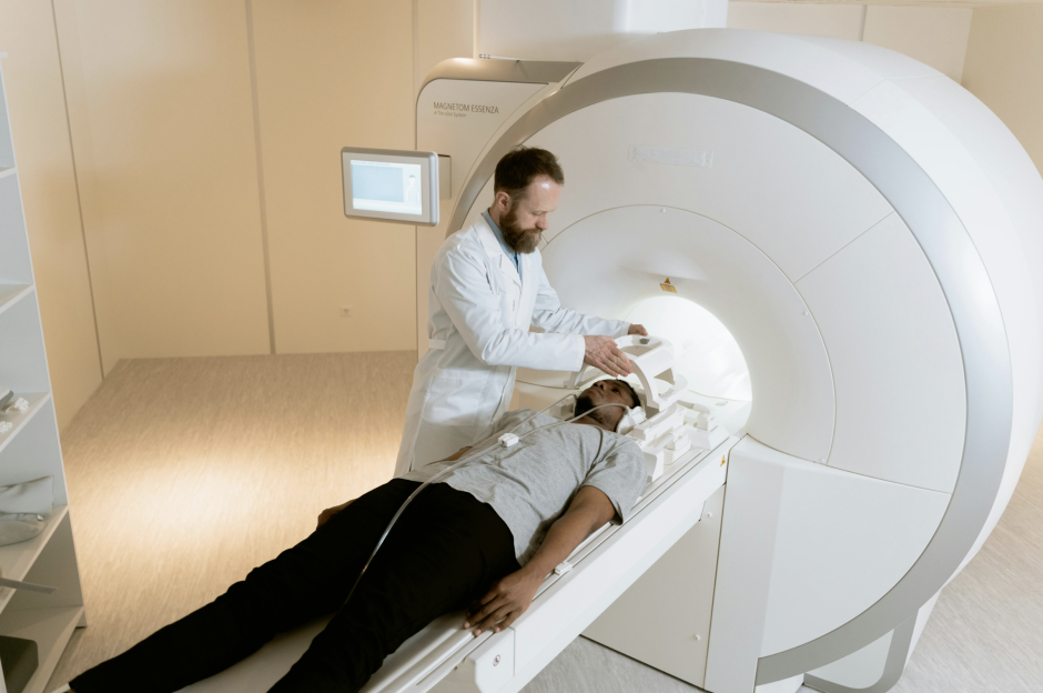
In emergency medicine, quick and accurate evaluation often starts with radiological imaging tools. Trauma patients may look stable but have serious internal injuries that doctors cannot see right away. High-impact events can damage bones, organs, or soft tissues without obvious external signs.
CT and MRI scans are used to uncover what the eye and hand might miss. But how do you decide which scan to use first in trauma care? When is MRI more valuable than CT in high-stakes injury situations? What role do these technologies play after the emergency room visit is over?
This article will explore how CT and MRI guide trauma care from initial response to patient recovery.
Choosing the Right Scan for the Situation
CT and MRI each serve different purposes in injury diagnosis. CT is fast and often the first scan used in trauma. It shows fractures, bleeding, and internal organ damage quickly and clearly. MRI takes longer but reveals soft tissue, brain, and spinal injuries better.
The Cleveland Clinic notes that some CT scans may take less than a minute. On the other hand, MRI scans can last 20 to 50 minutes. Patients must remain very still throughout the MRI to ensure accurate imaging results.
Doctors weigh time, patient condition, and injury type when choosing. Stable patients with nerve concerns often get an MRI after a CT.
Imaging choices shape surgery plans and help avoid further complications. Every case requires careful planning to match technology with patient needs.
What factors affect scan image quality?
Patient movement, body size, and metal in the body can degrade image clarity. Poor image quality may lead to missed diagnoses or repeat scans, delaying treatment decisions. Radiology teams may use positioning aids or contrast agents to improve imaging quality during complex evaluations.
Identifying Internal Injuries That Physical Exams Miss
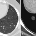
Physical exams cannot always detect serious damage after an accident. CT scans expose hidden injuries that need urgent treatment. MRI scans reveal soft tissue damage that might otherwise go unnoticed.
These tools make trauma care more accurate and thorough for patients. Internal bleeding or minor fractures may not show during basic checks. Head injuries that seem mild may reveal severe damage in imaging scans.
Medscape states that some signs that a mild head injury may actually be serious include vomiting, intense headache, memory loss, and loss of consciousness. People in this higher-risk group should receive a head CT scan for further evaluation. If the scan is clear, they may be discharged after a few hours of observation.
Early imaging reduces the risk of complications from delayed diagnosis. Doctors rely on these images to determine how urgent the case is. This ensures proper treatment from the very beginning of care.
What injuries most often go unnoticed early?
Small internal bleeds, ligament tears, or organ contusions can often be missed in initial checks. These injuries might not show symptoms until hours or days later, delaying treatment. Early imaging ensures such hidden issues are diagnosed before complications worsen the patient’s condition.
When Every Second Counts in Patient Care
Timing is crucial during emergency trauma evaluations involving severe injuries. As mentioned earlier, CT scans work quickly and deliver results in just a few minutes. This allows doctors to act before conditions become life-threatening for patients.
MRI offers more detail but usually follows CT in urgent cases. Emergency teams use CT first because fast information can save lives. Patients are stabilized based on initial scan results and then reassessed.
Follow-up MRI helps confirm precise damage to nerves or soft tissues. Doctors need fast and accurate tools to make the best decisions possible. Combining both methods improves care without wasting precious time.
What are the challenges in scanning unstable patients?
Unstable patients may struggle to lie still or be moved to scanning rooms safely. These conditions complicate imaging and may require sedation, special protocols, or mobile equipment. Balancing patient stabilization with imaging urgency remains one of trauma care’s most difficult decisions.
Communicating Injury Complexity Through Imaging
CT and MRI imaging help trauma care teams coordinate more effectively after a serious injury occurs. These scans provide diagnostic clarity and act as communication tools between physicians, surgeons, and support staff. In complex trauma cases, different specialists depend on imaging to understand the full scope of injuries. It allows the medical team to recognize the full impact of complicated injuries.
One such case was recently reported by KKTV involving a head-on crash in Colorado Springs. The incident happened on March 8, 2025, on the 2000 block of North Academy Boulevard. According to police, a vehicle traveling in the wrong direction caused a serious two-car collision. Both drivers were hospitalized with significant injuries requiring ongoing medical care and observation.
In scenarios like this, CT and MRI imaging help every provider understand the type and severity of trauma. Legal professionals may also be involved in cases like these to support the individuals affected. In this specific case, a personal injury lawyer in Colorado Springs could help review the circumstances and injuries.
According to Springs Law Group, these attorneys assist victims in recovering compensation for treatment, lost wages, and long-term medical support. Personal injury lawyers gather evidence, request documentation, and build cases based on medical findings and expert reports. Imaging studies often become a key part of this documentation due to their detail and accuracy.
These images help lawyers explain the impact of an injury and support the need for compensation. They also help prove that the trauma was caused by the specific incident under legal review. Overall, the importance of these scans goes far beyond the legal world and remains firmly rooted in clinical care. They guide both short-term interventions and long-term recovery decisions across all phases of care.
Can scan evidence support long-term disability claims?
Yes, detailed CT or MRI findings are crucial when filing for long-term disability benefits. These records show permanent damage or functional loss, which supports ongoing care and legal recognition. Imaging becomes a key part of the documentation required for government or private claims.
Improving Outcomes Through Integrated Imaging Strategy
Using CT and MRI together improves both diagnosis and patient recovery outcomes.
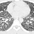
Cureus reports that combining CT with other diagnostic tools like MRI helps reduce unnecessary scans and improves clinical decisions. While MRI offers better long-term insight, CT remains essential for immediate evaluation in trauma cases. In acute traumatic brain injuries, CT supports faster triage and early treatment planning. MRI fills in details later, making both scans valuable at different stages of care.
Trauma teams compare both results to guide surgical and treatment decisions. Hospitals that integrate both scans report faster recovery times in patients. Better images lead to better choices during all stages of trauma care.
Moreover, new scan technology gives clearer images with shorter scan times than before. These advances support safer, more reliable trauma response strategies in hospitals.
Are scan results used in clinical trials?
Yes, trauma-focused clinical trials often use imaging to track treatment responses or compare protocols. CT and MRI provide objective markers for healing, inflammation, or deterioration over time. Imaging data also helps researchers develop evidence-based guidelines for emergency care and long-term outcomes.
Radiology tools like CT and MRI are essential when treating serious trauma and high-impact injuries. These scans reveal damage that physical exams might miss, helping doctors act quickly and accurately. CT is ideal for fast decisions in emergencies, while MRI captures soft tissue detail later.
Together, they guide surgical planning and make treatments more targeted and efficient. Imaging also improves communication between doctors, nurses, and other care teams involved in recovery. In many cases, scan results support legal claims and disability evaluations as well. Using both tools together improves patient recovery and enhances the overall delivery of trauma care.
Related posts:


Stay updated, free articles. Join our Telegram channel

Full access? Get Clinical Tree


