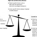
Contrast-enhanced ultrasonography (CEUS) is a rapidly evolving modality for imaging carotid artery disease and systemic atherosclerosis. CEUS coupled with diagnostic ultrasonography predicts the degree of carotid artery stenosis and is comparable with computed tomography and magnetic resonance angiography. This article reviews the literature on the evolving role of CEUS for the identification and characterization of carotid plaques with an emphasis on detection of intra-plaque neovascularization and related high-risk morphologic features notably present in symptomatic patients. CEUS carotid imaging may play a prominent additive role in risk stratifying patients and serve as a powerful tool for monitoring therapeutic interventions.
Detection of IPN using CEUS carotid artery imaging may be predictive of adverse cardiovascular events in patients with known coronary artery disease. If confirmed in larger clinical trials, CEUS carotid imaging may be useful for cardiovascular risk stratification by providing a marker of subclinical atherosclerosis.
Detection of IPN using CEUS carotid artery imaging may be predictive of adverse cardiovascular events in patients with known coronary artery disease. If confirmed in larger clinical trials, CEUS carotid imaging may be useful for cardiovascular risk stratification by providing a marker of subclinical atherosclerosis.
References
- 1. Beneficial effect of carotid endarterectomy in symptomatic patients with high-grade carotid stenosis. N Engl J Med 1991; 325: pp. 445-453
- 2. MRC European Carotid Surgery Trial: interim results for symptomatic patients with severe (70-99%) or with mild (0-29%) carotid stenosis. European Carotid Surgery Trialists’ Collaborative Group. Lancet 1991; 337: pp. 1235-1243
- 3. Brott T.G., Halperin J.L., Abbara S., et al: 2011 ASA/ACCF/AHA/AANN/AANS/ACR/ASNR/CNS/SAIP/SCAI/SIR/SNIS/SVM/SVS guideline on the management of patients with extracranial carotid and vertebral artery disease: a report of the American College of Cardiology Foundation/American Heart Association Task Force on Practice Guidelines, and the American Stroke Association, American Association of Neuroscience Nurses, American Association of Neurological Surgeons, American College of Radiology, American Society of Neuroradiology, Congress of Neurological Surgeons, Society of Atherosclerosis Imaging and Prevention, Society for Cardiovascular Angiography and Interventions, Society of Interventional Radiology, Society of NeuroInterventional Surgery, Society for Vascular Medicine, and Society for Vascular Surgery. J Am Coll Cardiol 2011; 57: pp. e16-e94
- 4. Pfister K., Rennert J., Greiner B., et al: Pre-surgical evaluation of ICA-stenosis using 3D power Doppler, 3D color coded Doppler sonography, 3D B-flow and contrast enhanced B-flow in correlation to CTA/MRA: first clinical results. Clin Hemorheol Microcirc 2009; 41: pp. 103-116
- 5. Cosgrove D.: Ultrasound contrast agents: an overview. Eur J Radiol 2006; 60: pp. 324-330
- 6. Ferrer J.M., Samso J.J., Serrando J.R., et al: Use of ultrasound contrast in the diagnosis of carotid artery occlusion. J Vasc Surg 2000; 31: pp. 736-741
- 7. Droste D.W., Jurgens R., Nabavi D.G., et al: Echocontrast-enhanced ultrasound of extracranial internal carotid artery high-grade stenosis and occlusion. Stroke 1999; 30: pp. 2302-2306
- 8. Köster W.: Endarteritis and arteritis. Berl Klin Wochenschr 1876; 13: pp. 454-455
- 9. Michel J.B., Martin-Ventura J.L., Nicoletti A., et al: Pathology of human plaque vulnerability: mechanisms and consequences of intraplaque haemorrhages. Atherosclerosis 2014; 234: pp. 311-319
- 10. Moulton K.S.: Plaque angiogenesis and atherosclerosis. Curr Atheroscler Rep 2001; 3: pp. 225-233
- 11. Shemirani B., and Crowe D.L.: Hypoxic induction of HIF-1alpha and VEGF expression in head and neck squamous cell carcinoma lines is mediated by stress activated protein kinases. Oral Oncol 2002; 38: pp. 251-257
- 12. Barger A.C., and Beeuwkes R.: Rupture of coronary vasa vasorum as a trigger of acute myocardial infarction. Am J Cardiol 1990; 66: pp. 41G-43G
- 13. Fleiner M., Kummer M., Mirlacher M., et al: Arterial neovascularization and inflammation in vulnerable patients: early and late signs of symptomatic atherosclerosis. Circulation 2004; 110: pp. 2843-2850
- 14. Dunmore B.J., McCarthy M.J., Naylor A.R., et al: Carotid plaque instability and ischemic symptoms are linked to immaturity of microvessels within plaques. J Vasc Surg 2007; 45: pp. 155-159
- 15. Fryer J.A., Myers P.C., and Appleberg M.: Carotid intraplaque hemorrhage: the significance of neovascularity. J Vasc Surg 1987; 6: pp. 341-349
- 16. Mofidi R., Crotty T.B., McCarthy P., et al: Association between plaque instability, angiogenesis and symptomatic carotid occlusive disease. Br J Surg 2001; 88: pp. 945-950
- 17. Moreno P.R., Purushothaman K.R., Fuster V., et al: Plaque neovascularization is increased in ruptured atherosclerotic lesions of human aorta: implications for plaque vulnerability. Circulation 2004; 110: pp. 2032-2038
- 18. Hellings W.E., Peeters W., Moll F.L., et al: Composition of carotid atherosclerotic plaque is associated with cardiovascular outcome: a prognostic study. Circulation 2010; 121: pp. 1941-1950
- 19. Howard D.P., van Lammeren G.W., Rothwell P.M., et al: Symptomatic carotid atherosclerotic disease: correlations between plaque composition and ipsilateral stroke risk. Stroke 2015; 46: pp. 182-189
- 20. Kono Y., Pinnell S.P., Sirlin C.B., et al: Carotid arteries: contrast-enhanced US angiography–preliminary clinical experience. Radiology 2004; 230: pp. 561-568
- 21. Faggioli G.L., Pini R., Mauro R., et al: Identification of carotid ‘vulnerable plaque’ by contrast-enhanced ultrasonography: correlation with plaque histology, symptoms and cerebral computed tomography. Eur J Vasc Endovasc Surg 2011; 41: pp. 238-248
- 22. Hammond C.J., McPherson S.J., Patel J.V., et al: Assessment of apparent internal carotid occlusion on ultrasound: prospective comparison of contrast-enhanced ultrasound, magnetic resonance angiography and digital subtraction angiography. Eur J Vasc Endovasc Surg 2008; 35: pp. 405-412
- 23. Baud J.M., Becker F., Maurizot A., et al: Contrast enhanced ultrasound can show symptomatic carotid lesions not visualized with magnetic resonance angiography. J Mal Vasc 2013; 38: pp. 385-391
- 24. Feinstein S.B.: The powerful microbubble: from bench to bedside, from intravascular indicator to therapeutic delivery system, and beyond. Am J Physiol Heart Circ Physiol 2004; 287: pp. H450-H457
- 25. Feinstein S.B.: Contrast ultrasound imaging of the carotid artery vasa vasorum and atherosclerotic plaque neovascularization. J Am Coll Cardiol 2006; 48: pp. 236-243
- 26. Vicenzini E., Giannoni M.F., Puccinelli F., et al: Detection of carotid adventitial vasa vasorum and plaque vascularization with ultrasound cadence contrast pulse sequencing technique and echo-contrast agent. Stroke 2007; 38: pp. 2841-2843
- 27. Coli S., Magnoni M., Sangiorgi G., et al: Contrast-enhanced ultrasound imaging of intraplaque neovascularization in carotid arteries: correlation with histology and plaque echogenicity. J Am Coll Cardiol 2008; 52: pp. 223-230
- 28. van den Oord S.C., Akkus Z., Bosch J.G., et al: Quantitative contrast-enhanced ultrasound of intraplaque neovascularization in patients with carotid atherosclerosis. Ultraschall Med 2015; 36: pp. 154-161
- 29. Saito K., Nagatsuka K., Ishibashi-Ueda H., et al: Contrast-enhanced ultrasound for the evaluation of neovascularization in atherosclerotic carotid artery plaques. Stroke 2014; 45: pp. 3073-3075
- 30. Ventura C.A., da Silva E.S., Cerri G.G., et al: Can contrast-enhanced ultrasound with second-generation contrast agents replace computed tomography angiography for distinguishing between occlusion and pseudo-occlusion of the internal carotid artery? Clinics (Sao Paulo) 2015; 70: pp. 1-6
- 31. ten Kate G.L., van Dijk A.C., van den Oord S.C., et al: Usefulness of contrast-enhanced ultrasound for detection of carotid plaque ulceration in patients with symptomatic carotid atherosclerosis. Am J Cardiol 2013; 112: pp. 292-298
- 32. Hjelmgren O., Johansson L., Prahl U., et al: A study of plaque vascularization and inflammation using quantitative contrast-enhanced US and PET/CT. Eur J Radiol 2014; 83: pp. 1184-1189
- 33. Shah F., Balan P., Weinberg M., et al: Contrast-enhanced ultrasound imaging of atherosclerotic carotid plaque neovascularization: a new surrogate marker of atherosclerosis? Vasc Med 2007; 12: pp. 291-297
- 34. Giannoni M.F., Vicenzini E., Citone M., et al: Contrast carotid ultrasound for the detection of unstable plaques with neoangiogenesis: a pilot study. Eur J Vasc Endovasc Surg 2009; 37: pp. 722-727
- 35. Varetto G., Gibello L., Bergamasco L., et al: Contrast enhanced ultrasound in atherosclerotic carotid artery disease. Int Angiol 2012; 31: pp. 565-571
- 36. Shalhoub J., Monaco C., Owen D.R., et al: Late-phase contrast-enhanced ultrasound reflects biological features of instability in human carotid atherosclerosis. Stroke 2011; 42: pp. 3634-3636
- 37. Hoogi A., Adam D., Hoffman A., et al: Carotid plaque vulnerability: quantification of neovascularization on contrast-enhanced ultrasound with histopathologic correlation. AJR Am J Roentgenol 2011; 196: pp. 431-436
- 38. Vavuranakis M., Sigala F., Vrachatis D.A., et al: Quantitative analysis of carotid plaque vasa vasorum by CEUS and correlation with histology after endarterectomy. Vasa 2013; 42: pp. 184-195
- 39. Li C., He W., Guo D., et al: Quantification of carotid plaque neovascularization using contrast-enhanced ultrasound with histopathologic validation. Ultrasound Med Biol 2014; 40: pp. 1827-1833
- 40. Müller H.F., Viaccoz A., Kuzmanovic I., et al: Contrast-enhanced ultrasound imaging of carotid plaque neo-vascularization: accuracy of visual analysis. Ultrasound Med Biol 2014; 40: pp. 18-24
- 41. Iezzi R., Petrone G., Ferrante A., et al: The role of contrast-enhanced ultrasound (CEUS) in visualizing atherosclerotic carotid plaque vulnerability: Which injection protocol? Which scanning technique? Eur J Radiol 2015; 84: pp. 865-871
- 42. Zhou Y., Xing Y., Li Y., et al: An assessment of the vulnerability of carotid plaques: a comparative study between intraplaque neovascularization and plaque echogenicity. BMC Med Imaging 2013; 13: pp. 13
- 43. Shao A., Dong X., Zhou J., et al: Comparison of carotid artery endarterectomy and carotid artery stenting in patients with atherosclerotic carotid stenosis. J Craniofac Surg 2014; 25: pp. 1441-1447
- 44. Varetto G., Gibello L., Faletti R., et al: Contrast-enhanced ultrasound to predict the risk of microembolization during carotid artery stenting. Radiol Med 2015; undefined:
- 45. Clevert D.A., Sommer W.H., Helck A., et al: Duplex and contrast enhanced ultrasound (CEUS) in evaluation of in-stent restenosis after carotid stenting. Clin Hemorheol Microcirc 2011; 48: pp. 199-208
- 46. Sampson U.K., Harrell F.E., Fazio S., et al: Carotid adventitial vasa vasorum and intima-media thickness in a primary prevention population. Echocardiography 2015; 32: pp. 264-270
- 47. Xiong L., Li P., and Zhao B.W.: Evaluation of carotid plaque neovascularization in patients with diabetes mellitus by contrast-enhanced ultrasonography. J Huazhong Univ Sci Technolog Med Sci 2014; 34: pp. 29-32
- 48. van den Oord S.C., Akkus Z., Renaud G., et al: Assessment of carotid atherosclerosis, intraplaque neovascularization, and plaque ulceration using quantitative contrast-enhanced ultrasound in asymptomatic patients with diabetes mellitus. Eur Heart J Cardiovasc Imaging 2014; 15: pp. 1213-1218
- 49. van den Oord S.C., Akkus Z., Roeters van Lennep J.E., et al: Assessment of subclinical atherosclerosis and intraplaque neovascularization using quantitative contrast-enhanced ultrasound in patients with familial hypercholesterolemia. Atherosclerosis 2013; 231: pp. 107-113
- 50. Arcidiacono M.V., Rubinat E., Borras M., et al: Left carotid adventitial vasa vasorum signal correlates directly with age and with left carotid intima-media thickness in individuals without atheromatous risk factors. Cardiovasc Ultrasound 2015; 13: pp. 20
- 51. van den Oord S.C., ten Kate G.L., Sijbrands E.J., et al: Effect of carotid plaque screening using contrast-enhanced ultrasound on cardiovascular risk stratification. Am J Cardiol 2013; 111: pp. 754-759
- 52. Staub D., Patel M.B., Tibrewala A., et al: Vasa vasorum and plaque neovascularization on contrast-enhanced carotid ultrasound imaging correlates with cardiovascular disease and past cardiovascular events. Stroke 2010; 41: pp. 41-47
- 53. Huang P.T., Chen C.C., Aronow W.S., et al: Assessment of neovascularization within carotid plaques in patients with ischemic stroke. World J Cardiol 2010; 2: pp. 89-97
- 54. Zhu Y., Deng Y.B., Liu Y.N., et al: Use of carotid plaque neovascularization at contrast-enhanced US to predict coronary events in patients with coronary artery disease. Radiology 2013; 268: pp. 54-60
- 55. Deyama J., Nakamura T., Takishima I., et al: Contrast-enhanced ultrasound imaging of carotid plaque neovascularization is useful for identifying high-risk patients with coronary artery disease. Circ J 2013; 77: pp. 1499-1507
- 56. Yoshida J., Ohmori K., Takeuchi H., et al: Treatment of ischemic limbs based on local recruitment of vascular endothelial growth factor-producing inflammatory cells with ultrasonic microbubble destruction. J Am Coll Cardiol 2005; 46: pp. 899-905
- 57. ten Kate G.L., Renaud G.G., Akkus Z., et al: Far-wall pseudoenhancement during contrast-enhanced ultrasound of the carotid arteries: clinical description and in vitro reproduction. Ultrasound Med Biol 2012; 38: pp. 593-600
- 58. Lantheus Medical Imaging. Definity (perflutren lipid microsphere) full prescribing information (product insert). Lantheus Medical Imaging website (US). Available at: http://www.definityimaging.com/pdf/DEFINITY%20Prescribing%20Information%20515987-0413.pdf 2013;515987-0413. Accessed May 1, 2015.
- 59. GE Healthcare. Optison (perflutren protein type-A microspheres injectable suspension, USP) full prescribing information (product insert). GE Healthcare website (US). Available at: http://www.optisonimaging.com/us/wp-content/uploads/2014/10/Updated-Optison-PI-10-21-14.pdf 2013;OPT-1F-OSLO. Accessed May 1, 2015.
Related posts:
 Three-Dimensional Carotid Plaque MR Imaging
Incorporating Carotid Plaque Imaging into Routine Clinical Carotid Magnetic Resonance Angiography
FDG PET/CT Imaging of Carotid Atherosclerosis
Clinical Perspective of Carotid Plaque Imaging
Three-Dimensional Carotid Plaque MR Imaging
Incorporating Carotid Plaque Imaging into Routine Clinical Carotid Magnetic Resonance Angiography
FDG PET/CT Imaging of Carotid Atherosclerosis
Clinical Perspective of Carotid Plaque Imaging
 Plaque Imaging to Decide on Optimal Treatment
 Low-Grade Carotid Stenosis
Stay updated, free articles. Join our Telegram channel

Full access? Get Clinical Tree


