Figure 31.1.1
Transvaginal ultrasound-guided aspiration of a unilocular cyst. A: Transvaginal view of the right adnexa demonstrates a simple unilocular cyst (arrows). There is a needle guide on the transducer, and the guide marks on the image indicate the path that will be taken by a needle inserted through the guide. B: A needle (arrowheads) has been inserted to drain the cyst.
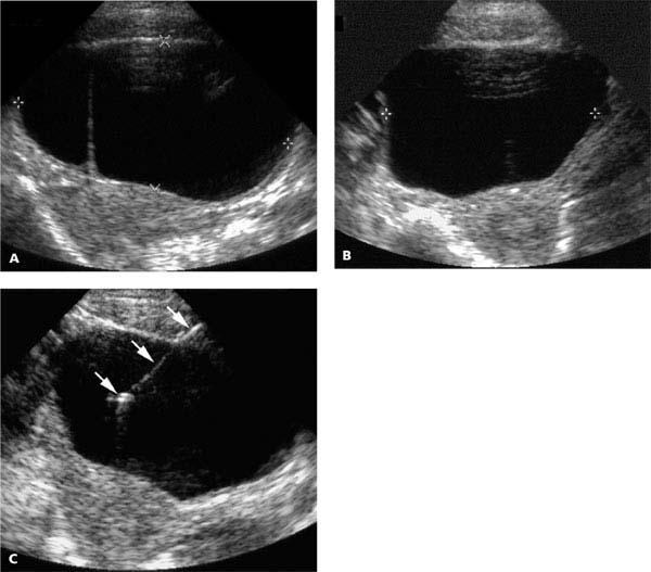
Figure 31.1.2
Transabdominal ultrasound-guided aspiration of a multilocular cyst. A: Sagittal and (B) transverse transabdominal views of the right adnexa demonstrate a septated cyst (calipers). C: A needle (arrows) has been inserted in the larger locule under freehand transabdominal ultrasound guidance, without the use of a needle guide attachment.
31.2. Oocyte Retrieval
Description and Clinical Features
When a woman undergoes in vitro fertilization, she takes medication to stimulate her ovaries to produce multiple follicles. Many of these follicles contain oocytes (i.e., eggs). The oocytes are retrieved using ultrasound-guided aspiration of the follicles. The oocytes are fertilized by incubating them with the father’s sperm, and the resultant embryos are then transferred into the woman’s uterus.
Sonography
Egg retrieval is usually performed under transvaginal ultrasound guidance. In some cases, when the ovaries are located high in the pelvis and close to the anterior abdominal wall, transabdominal guidance may be the better choice (Figure 31.2.1). With either guidance approach, the needle is advanced into each follicle and fluid is aspirated. The fluid is then examined for the presence of an oocyte.
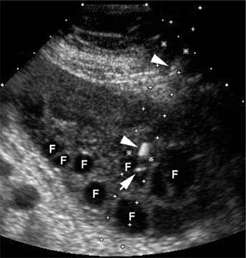
Figure 31.2.1
Transabdominal oocyte retrieval. There are multiple follicles (F) in this ovary, a result of follicular-stimulating medication. A needle (arrowheads) has been inserted into the ovary, with its tip (arrow) in one of the follicles.
31.3. Ultrasound-Guided Uterine Instrumentation Through the Cervix
Description and Clinical Features
Uterine instrumentation procedures, such as dilation and curettage (D&C) and office endometrial biopsy, are generally performed blindly. There are several situations in which ultrasound guidance is valuable, including the following:
Difficulty passing an instrument through the cervical canal (e.g., cervical stenosis or sharply ante- or retroverted uterus).
Ensuring complete evacuation of abnormal uterine cavity contents.
Assisting in the removal of an intrauterine device.
Sonography
Guidance of uterine instrumentation is performed using transabdominal ultrasound. It is done through a completely or partially filled urinary bladder.

Figure 31.3.1
Ultrasound-guided dilation and curettage. Sagittal transabdominal view of the uterus demonstrates a suction device (arrows) within the uterine cavity after it was passed through the cervix using sonographic guidance.
When a D&C or other procedure is hindered because of the inability to pass an instrument through the cervix, ultrasound can assist by determining the cervical orientation and directing the instrument along the long axis of the cervix. If necessary, the cervical orientation can be modified by filling or emptying the bladder. Once the instrument and cervix are properly aligned, forward pressure on the device will usually succeed in getting it through the cervical canal (Figure 31.3.1).
When suctioning purulent material, retained products of conception, or other contents from the uterine cavity, ultrasound monitoring is useful to determine when the cavity is empty (Figure 31.3.2). This ensures that the drainage procedure is not terminated prematurely when there is still material left in the cavity, nor prolonged unnecessarily after the cavity is empty.

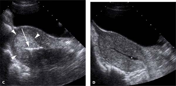
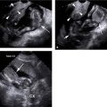
Figure 31.3.2
Ultrasound-guided evacuation of retained products of conception. A: Sagittal transabdominal image of the uterus in a woman who suffered a recent miscarriage demonstrates a soft-tissue mass (arrowheads) in the uterine cavity. B: Transverse transabdominal view, with blood flow identified within the mass on color Doppler. The mass represents retained products of conception. C: A suction device (arrow) lies within the retained products of conception (arrowheads). D: At the end of the procedure, there is a small amount of fluid (*) in the uterine cavity, but no abnormal tissue remains.
Related posts:


Stay updated, free articles. Join our Telegram channel

Full access? Get Clinical Tree


