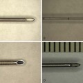
15 Three-Dimensional Ultrasound
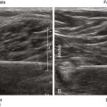
There are several reports of the use of three- or four-dimensional imaging to image nerves and guide regional blocks.1–3 The complexity of the surrounding echoes in musculoskeletal tissue can make rendering clear three-dimensional images challenging. Injected anechoic fluid can improve the interface for three-dimensional imaging of the nerve surface. Rendered volumes are often shown with sepia coloring to improve contrast resolution.
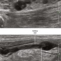
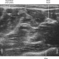
One potential advantage of three-dimensional imaging is to avoid partial line-ups of the block needle that can occur with two-dimensional in-plane technique. Because line-up is not necessary, performance time and accuracy of the procedure would benefit. One study found that the use of higher-dimensional imaging improved needle tip identification.4 However, another study found that multiplanar reformatted displays improved needle conspicuity compared with volume-rendered displays.5 Serious considerations for this developing technology balance obtaining more useful information with unnecessary distraction. Interventional procedure times tend to be longer with this technology.6
Related posts:


Stay updated, free articles. Join our Telegram channel

Full access? Get Clinical Tree


