Overview
Location
Thyroid Gland
• Located in the lower, anterior portion of the neck, anterior to the trachea and inferior to the larynx.
• Has bilateral lobes that lie on each side of the trachea. They are bordered laterally by the common carotid artery and internal jugular vein.
• The isthmus of the thyroid gland unites the lobes at the level of the second, third, and fourth tracheal rings.
• Lies between the anterior strap muscles: (sternocleidomastoid, sternothyroid, and sternohyoid muscles) and the posterior longus colli muscle and minor neurovascular bundle (consisting of the inferior thyroid artery and recurrent laryngeal nerve).
Parathyroid Glands
• The 4 glands lie between the posterior aspect of the thyroid gland and the longus colli muscle.
• The 2 superior parathyroid glands are situated slightly more medial than the 2 inferior parathyroid glands.
• Typically, the parathyroid glands are symmetric in position.
• Lie deep to the cricoid cartilage, midway between the Adam’s apple (apex of thyroid cartilage) and the suprasternal notch.
Anatomy
Thyroid Gland
• A superficial, butterfly-shaped gland that is variable in size but weighs approximately 25 to 35 g.
• The size and shape of the thyroid gland is greatly influenced by body habitus and gender, therefore, normal thyroid gland measurements have a wide range of variability:
• Tall, thin individuals have elongated lateral lobes that can measure up to 8 cm long.
• Short, obese people’s lateral lobes are 5 cm or less.
• The thyroid gland is normally larger in women and becomes enlarged during pregnancy.
• The average, normal adult thyroid gland is approximately 4-6 cm long, 1.3 to 1.8 cm anteroposteriorly, and 3 cm at its greatest width. The anteroposterior measurement of the isthmus is approximately 2 to 6 cm.
• In newborns and children, the gland is approximately 2 to 3 cm long, 0.2 to 1.2 cm anteroposteriorly, and 1.5 cm at its greatest width.
• The thyroid is highly vascular; its blood supply consists of paired superior and inferior arteries and veins, and often, middle thyroid veins.
Parathyroid Glands
• The oval or bean shaped parathyroid glands are approximately 5 to 7 mm long, 1 to 2 mm thick, and 3 to 4 mm wide.
• Parathyroid glands are supplied blood by separate small branches of the inferior and superior thyroid arteries. Venous drainage is via the superior, inferior, and middle thyroid veins.
Physiology
Thyroid Gland
• An endocrine gland (ductless gland that secretes hormones directly into the bloodstream) that is responsible for secreting the hormones thyroxine (T4) (regulates metabolism), triiodothyronine (T3) (regulates metabolism), and calcitonin (decreases blood calcium levels, preventing hypercalcemia) that assist in regulating the metabolic rate and the metabolism of lipids, proteins, and carbohydrates and in the absorption of calcium from the blood for storage in the bones.
Parathyroid Glands
• Endocrine glands that secrete the hormone parathormone (PTH) which acts as the antagonist for calcitonin and controls the calcium level in the bloodstream.
Sonographic Appearance
Thyroid Gland
• Appears homogeneous and moderately echogenic. It is similar in appearance to normal liver and testes parenchyma and hyperechoic compared to adjacent musculature.
• For appearance’s sake, the long axis of the thyroid gland is situated horizontally in the neck. Therefore, longitudinal and long axis views of the entire gland would be visualized in transverse scanning planes. However, for diagnostic purposes the thyroid’s right and left lobes and the isthmus that connects them are examined separately.
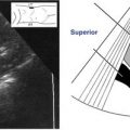
• Each lobe is oriented vertically in the neck; therefore, the long axes are visualized in sagittal scanning planes. The long sections of the lobes appear sandwiched between the hypoechoic strap muscles anteriorly and longus colli muscles posteriorly (Figure 16-1).
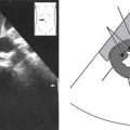
• Axial sections of the right and left lobes are seen extending laterally from each side of the isthmus to axial sections of the common carotid arteries and internal jugular veins, which appear as anechoic round or oval structures with bright walls. In transverse scanning plane images showing axial sections of the thyroid lobes, adjacent neck structures such as the vagus nerve, recurrent laryngeal nerve, the esophagus, and various muscles are routinely identified (Figure 16-2).
▪ The vagus nerve (part of the major neurovascular bundle) is visualized as a hypoechoic dot posterolateral to the thyroid lobe, usually between the carotid artery and jugular vein.
▪ The recurrent laryngeal nerve (part of the minor neurovascular bundle) is best identified as an echogenic rim located between the trachea, esophagus, and left posterior thyroid lobe.


▪ The esophagus (part of the alimentary canal that connects the throat to the stomach) is seen just to the left of the midline of the neck, posterior to the left thyroid lobe, between the trachea (large membranous tube reinforced by rings of cartilage, extending from the larynx to the bronchial tubes and conveying air to and from the lungs) and longus colli muscle. Its axial section appears round to oval; the outer wall is hypoechoic relative to the bright appearance of the inner lumen wall. The lumen can, however, appear anechoic or with a centrally located, moderately echogenic line or dot representing mucosa. The longus colli muscle extends laterally to lie posterior to the common carotid artery.
▪ Neck muscles appear distinctively hypoechoic compared to the thyroid gland. Anteriorly, the strap muscles (sternohyoid, sternothyroid, omohyoid, and sternocleidomastoid) are easily distinguished as is the longus colli muscle posteriorly.
• The long axis of the isthmus is situated horizontally and viewed from a transverse scanning plane at the midline of the neck, just anterior to the bright cartilaginous rings of the trachea.
• Branches of the inferior and superior thyroid arteries and veins appear as 1 to 2 mm anechoic structures with thin, bright walls. Color Doppler can be helpful in differentiating intra- and extrathyroidal vessels.
Parathyroid Glands
• By using newer high frequency transducers, normal parathyroid glands are occasionally visualized sonographically, especially in young children. When identified, they appear as a flat, hypoechoic structure lying between the thyroid gland anteriorly and longus colli muscle posteriorly.
Color Flow Doppler Characteristics
• Utilize color Doppler to differentiate vascular from nonvascular structures.
• Adjust color Doppler parameters to detect normal or increased flow in the thyroid gland.
• Color Doppler shows increased vascularity within and in the periphery of autonomous functioning adenomas (a benign tumor formed from glands) and thyroid cancer.
• Utilize color Doppler to follow the superior or inferior thyroid arteries to locate parathyroid adenomas.
Normal Variants
Thyroid Gland
• Pyramidal lobe: Triangular-shaped, superior extension of the isthmus. Present in 15% to 30% of thyroid glands. Variable in size and extends more often to the left side. Parenchyma appears the same as the normal thyroid.
Related posts:
Stay updated, free articles. Join our Telegram channel

Full access? Get Clinical Tree


