and Zdeněk Fryšák1
(1)
Department of Internal Medicine III – Nephrology, Rheumatology and Endocrinology, Faculty of Medicine and Dentistry, Palacky University Olomouc and University Hospital Olomouc, Olomouc, Czech Republic
Keywords
Pure cystsComplex cysts“Comet tails”DebrisSepta7.1 Essential Facts
At sonography, 15–25% of solitary thyroid nodules are found to be cystic or predominantly cystic [1, 2].
Based on the ratio of the liquid portion in nodule, nodules are classified as: mixed predominantly cystic nodule (liquid portion >50% but ≤90% of the nodule volume) or cystic nodule (liquid portion >90% of the nodule volume) [3].
As thyroid cysts are considered nodules with a liquid component of more than 60% volume; this strict definition is used especially for percutaneous ethanol injection therapy (PEIT) studies; see more in Chap. 23 [4, 5].
Thyroid cysts are divided into pure cysts – without internal septa (Fig. 7.1aa) or complex/polyconcamerated cysts – with one or more internal septa (Figs. 7.6aa and 7.8aa) [6].
In pure cysts the fluid content is colloid only, occasionally with the condensed colloid proteins, while in complex cysts the fluid component may also be the result of degeneration or hemorrhage [6].
Pure cystic lesions are always thought to be benign; on the contrary, in complex cysts the solid component may represent a 3% risk of malignancy [7].
Primary thyroid cysts, although rare (<2% of thyroid lesions), are highly likely to be benign [8].
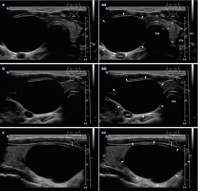
Fig. 7.1
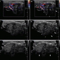
(aa) A 43-year-old woman with a solitary large pure cyst (arrowheads), size 42 × 31 × 26 mm and volume 17 mL in the RL. US overall view: round shape; homogeneously anechoic contents; smooth wall; Tvol 28 mL, asymmetry—RL 24 mL and LL 4 mL; transverse. (bb) Detail of large pure cyst (arrowheads): ovoid shape; anechoic contents; smooth wall; posterior acoustic enhancement; transverse. (cc) Detail of large pure cyst (arrowheads): ovoid shape; anechoic contents; smooth wall; posterior acoustic enhancement; longitudinal
7.2 US Features of Thyroid Cysts [6]
Pure cysts (Figs. 7.1aa and 7.2aa):
Smooth wall, possibly with focal small solid hyperechoic component (Fig. 7.2aa).
Fluid usually appears homogeneously anechoic.
Posterior acoustic enhancement (Fig. 7.1cc).
Bright hyperechoic reverberation artifacts so-called “comet tails” could be found in the fluid content (condensed colloid proteins).
A single “comet tail” artifact within a small cyst is usually called “cat’s eye artifact” (Fig. 7.3bb, cc).
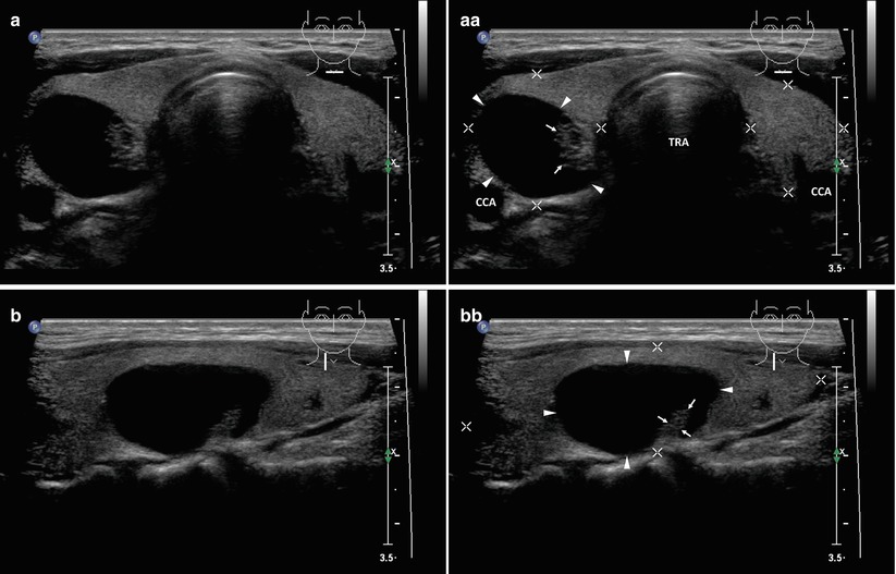
Fig. 7.2
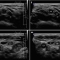
(aa) A 38-year-old woman with a solitary small pure cyst (arrowheads), size 25 × 20 × 14 mm and volume 3.5 mL in the RL: ovoid shape; homogeneously anechoic contents; smooth wall with focal solid hyperechoic component (arrows); Tvol 19 mL, RL 12 mL, and LL 7 mL; transverse. (bb) Detail of small pure cyst (arrowheads): ovoid shape; anechoic contents; smooth wall with focal solid hyperechoic component (arrows) as a polyp extending from the wall; longitudinal
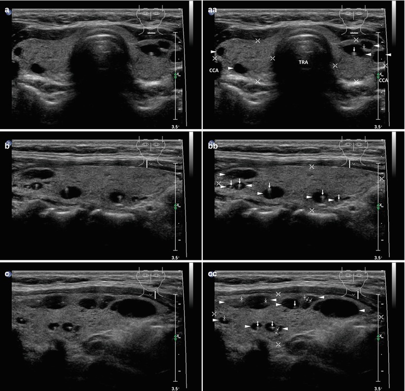
Fig. 7.3
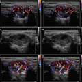
(aa) A 44-year-old woman with multiple tiny cysts (arrowheads) sized from 4 to 7 mm bilaterally: round or oval shape; anechoic contents with single comet tails (colloid clot)—“cat’s eye artifacts” (arrows); Tvol 12 mL, RL 6 mL, and LL 6 mL; transverse. (bb) Detail of the RL with multiple tiny cyst (arrowheads): “cat’s eye artifacts” (arrows); sporadic tiny colloid clot without tails (open arrows); longitudinal. (cc) Detail of the LL with multiple tiny cyst (arrowheads): “cat’s eye artifacts” (arrows); sporadic tiny colloid clot without tails (open arrows); longitudinal
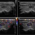
Complex cysts (Figs. 7.4aa and 7.8aa):