and Vincent L. Sorrell2
(1)
Division of Nuclear Medicine and Molecular Imaging Department of Radiology, University of Kentucky, Lexington, Kentucky, USA
(2)
Division of Cardiovascular Medicine Department of Internal Medicine Gill Heart Institute, University of Kentucky, Lexington, Kentucky, USA
Electronic supplementary material
The online version of this chapter (doi: 10.1007/978-3-319-25436-4_4) contains supplementary material, which is available to authorized users.
MPI radiopharmaceuticals normally localize in the thyroid gland. Benign and malignant thyroid conditions may accumulate these radiopharmaceuticals. Accordingly, apart from MPI, they have been used diagnostically for the evaluation of non-iodine-avid thyroid cancer and for the evaluation of thyroid nodules (Seabold et al. 1999; Sundram and Mack 1995). “Hot” lesions tend to be more easily recognized, but, at times, the degree of localization may actually be less than the surrounding or adjacent normal thyroid tissue, and the lesion may appear relatively “cold.”
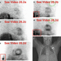
Situated in the lower neck, the thyroid gland is often included at the top of the imaging field-of-view during routine SPECT MPI (Fig. 4.1), particularly when using large detectors. The degree of uptake is usually relatively faint, but images can be manipulated to enhance its visualization (Fig. 4.1b, d). Review of the raw projection images can lead to identification of a variety of underlying thyroid conditions (Williams et al. 2003). Table 4.1 lists potential “hot” and “cold” imaging findings related to the thyroid gland.


Fig. 4.1
Normal thyroid gland at top of field-of-view. Normal thyroid activity can be identified on rest (a, b) and stress (c–e) images.(a) Rest raw projection images (Video 4.1a, frame 1) at usual contrast setting for the heart, 99mTc sestamibi. (b) Rest raw projection images (Video 4.1b, frame 1) with enhanced contrast for the thyroid gland, 99mTc sestamibi. (c) Stress raw projection images (Video 4.1c, frame 1) at usual contrast setting for the heart, 99mTc sestamibi. (d) Stress raw projection images (Video 4.1d, frame 1) with enhanced contrast for the thyroid gland, 99mTc sestamibi. (e) Stress raw projection image (Video 4.1e, frame 16) with enhanced contrast for the thyroid gland, 99mTc sestamibi, bilobed thyroid gland (yellow box), heart for anatomic reference only (blue lines)
Table 4.1
Differential diagnosis of “hot” and “cold” imaging findings related to the thyroid gland
Organ system | “Hot” finding | “Cold” finding | References |
|---|---|---|---|
Thyroid gland | Multinodular goiter Diffuse toxic goiter Substernal goiter Nodule/neoplasm, benign Nodule/neoplasm, malignant Lymphoma Free 99mTc pertechnetate | Cyst Neoplasm, primary
Stay updated, free articles. Join our Telegram channel
Full access? Get Clinical Tree
 Get Clinical Tree app for offline access

|

