Training and Program Development
 STEPS TO ESTABLISHING A POINT-OF-CARE ULTRASOUND PROGRAM
 DETERMINE TYPE OF EXAMINATIONS TO BE PERFORMED
 DEVELOP A PROGRAM IMPLEMENTATION PLAN
 SELECTING THE ULTRASOUND DIRECTOR(S)
 OBTAIN HOSPITAL APPROVAL OF THE PROGRAM
 ACQUIRE AN ULTRASOUND MACHINE
 TRAINING THE ULTRASOUND DIRECTOR
 TRAINING THE GROUP
 PERFORM PROBLEM SOLVING
 REGISTERED DIAGNOSTIC MEDICAL SONOGRAPHER
 ELECTIVE TRAINING
 FELLOWSHIP TRAINING
 COST OF A POINT-OF-CARE ULTRASOUND PROGRAM
 CODING AND REIMBURSEMENT
Establishing a training program in point-of-care ultrasound is an exciting and rewarding experience. The impact of ultrasound on the clinical practice of medicine becomes so clear that many clinicians, after acquiring basic ultrasound skills, wonder how they got along without this technology. This chapter outlines the process for developing a point-of-care ultrasound training program and addresses the common questions encountered when starting a new program.
Point-of-care ultrasound examinations are performed in real time at the bedside by clinicians to answer specific questions in order to expedite care and improve patient care. These studies are not intended to provide comprehensive surveys of anatomical areas nor are they mere extensions of the physical examination.1 Point-of-care ultrasound provides imaging to rule in or rule out specific disease entities for which timely treatment is crucial (e.g., ruptured abdominal aortic aneurysm, ruptured ectopic pregnancy, and cardiac tamponade) or for whom invasive intervention could be especially unsafe (e.g., paracentesis, abscess drainages, and foreign body removal). As such, these studies require the highest levels of competence, accuracy, and clinical acumen.
 STEPS TO ESTABLISHING A POINT-OF-CARE ULTRASOUND PROGRAM
In order to establish a high-quality point-of-care ultrasound program, ultrasound directors must:
1. Determine type of examinations to be performed.
2. Develop a program implementation plan.
3. Obtain leadership approval of the implementation plan.
4. Acquire an ultrasound machine.
5. Train the group.
6. Incentivize group members to complete training and credentialing.
7. Perform problem solving, quality assurance, and ongoing training.
A critical factor in the timely implementation and success of an ultrasound program is to have the full support and active assistance of the department leadership. Also, appointing a dedicated ultrasound director with protected time is the best approach because each step in program implementation is very time intensive.
 DETERMINE TYPE OF EXAMINATIONS TO BE PERFORMED
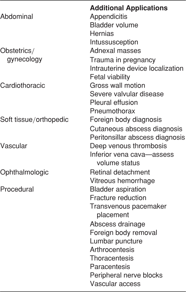
Ultrasound use continues to expand along with technological advances and improvement in individual operator expertise. It makes sense to start with applications that will get the most use in a particular practice setting. For example, in a small community hospital, evaluation of cardiac arrest may be more pertinent than trauma evaluations. Likewise, in centers without 24-hour ultrasound services, it may be important to learn ultrasound for the evaluation of ectopic pregnancy and cholecystitis. We recommend starting with applications unique to the ED for which timely diagnosis and intervention are critical. This includes the focused assessment with sonography for trauma (FAST) examination, evaluation of cardiac arrest states, and evaluation of hypotension. In addition, procedural applications, such as intravenous line placement, paracentesis, thoracentesis, and abscess localization and drainage, are becoming standard of care.2 Training in emergency medicine residency programs, however, should cover all of the primary applications and include exposure to emerging applications (Tables 1-1 and 1-2).
 TABLE 1-1. CORE EMERGENCY ULTRASOUND APPLICATIONS
Trauma
Intrauterine pregnancy
Abdominal aortic aneurysm
Cardiac
Biliary
Urinary tract
Deep venous thrombosis
Soft tissue/musculoskeletal
Ocular
Thoracic
Procedural guidance
Adapted from American College of Emergency Physicians: Emergency ultrasound guidelines. Ann Emerg Med 53:550–570, 2009.
 TABLE 1-2. EMERGENCY ULTRASOUND—ADDITIONAL APPLICATIONS
A new point-of-care ultrasound program should strive to identify all of the ultrasound examinations that are of interest, both now and in the future. Making this decision early allows the program to seek leadership approval for all of these examinations from the beginning instead of having to apply for additional approval later. It also allows the program to define equipment needs prior to initial equipment purchase. Otherwise, additional purchases may have to be made as new applications are brought online.
Some medical centers may choose to focus on just one or two ultrasound applications at a time. This allows everyone time to concentrate their efforts on becoming proficient with each application and to learn the technical pitfalls inherent in those particular applications. By keeping the entire training group on the same application(s), the ultrasound director can focus quality improvement (QI) efforts on those specific applications, using reviews of specific cases to educate and train everyone involved. After quality performance levels are achieved, new applications can be introduced in a similar fashion one at a time until all of the applications are taught. In our experience, this is the most effective means for starting a program and allows for a safe and effective implementation of ultrasound into clinical care.
Since point-of-care ultrasound focuses on critical questions or interventions, rather than requiring “minimal training,” the clinicians actually require the best training and highest standards of competence. In our experience, this is best accomplished by focused and appropriately directed program implementation and training.
 DEVELOP A PROGRAM IMPLEMENTATION PLAN
The program implementation plan defines all aspects of the ultrasound training program. The plan guides the ultrasound director through all the administrative and teaching aspects of the program and serves as a reference for requirements in training. The most straightforward way to develop a plan is to model it after another group or institution’s plan, adapting it for the local clinical and political environment. Many residency programs are willing to share their program plans. The program plan should, at a minimum, include the following elements:
 Definition of specific privileges.
 Training and credentialing.
 Method of recording results.
 Performance improvement plan.
 CME requirements.
DEFINITION OF SPECIFIC PRIVILEGES
This section defines exactly how focused ultrasound examinations will be used. For example, a specific privilege may be: “Documentation of free abdominal fluid in trauma patients.” This could also be shortened to “Documentation of free abdominal fluid” if there is an additional desire to diagnose ascites. Some institutions allow graduated privileges, meaning that clinicians can do more with their ultrasound examinations as they gain experience (Table 1-3). This approach is beneficial in that it allows earlier implementation of point-of-care ultrasound into patient care while establishing ongoing quality control parameters. Earlier implementation helps maintain momentum in the training program as clinicians see the benefits of their training sooner; however, graduated privileges create a more complex training program.
 TABLE 1-3. EXAMPLES OF LEVELS OF PROFICIENCY
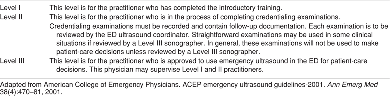
Privileges and training should be constructed toward the identification of specific findings (e.g., presence or absence of gallstones) and not toward the general evaluation of disease processes or anatomic structures (e.g., evaluation for “cardiac disease” or “right upper quadrant abdominal pain”) consistent with existing guidelines. A useful resource for defining a particular examination is the Emergency Ultrasound Imaging Criteria Compendium.3
TRAINING AND CREDENTIALING
Training
Multiple guidelines for training in ultrasound have been published.3–8 The American College of Radiology (ACR) and the American Institute of Ultrasound in Medicine (AIUM) have established guidelines4,5 for learning comprehensive examinations, but have not published guidelines for point-of-care ultrasound. In 1994, a model curriculum for training in emergency ultrasound was published by the Society for Academic Emergency Medicine (SAEM) and subsequently modified as new evidence and experience emerged.6 In 2001, the American College of Emergency Physicians (ACEP) published initial emergency ultrasound guidelines outlining utilization for six “primary applications” plus use for procedural guidance. The Council of Emergency Medicine Residency Directors (CORD) outlined ultrasound training standards for emergency medicine residency programs in 2008. They listed four primary applications (FAST, emergent cardiac, abdominal aortic aneurysm, intrauterine pregnancy) plus procedural guidance as the minimum skill set, but highly recommended training in at least six additional applications. ACEP published revised guidelines on ultrasound training that have expanded the list of “Core Applications” to a total of 11 applications1 (Table 1-1). Although this expanded list provides support for additional uses, it is recognized that not all facilities may have the need to utilize all of these applications in their specific clinical practice setting. Specific didactic and experiential training criteria are outlined with a minimum of 25 training cases documented for each core application of interest. The ACEP guidelines are recognized as the current standard for training in point-of-care ultrasound.7,8 Ultrasound directors still have some degree of latitude in designing their individual programs, but should consider CORD as well as ACEP guidelines when designing minimum training criteria. A program should also include minimum requirements for each of the following areas:
 Minimum overall didactic hours.
 Minimum overall didactic content.
 Minimum number of overall ultrasound examinations performed.
 Minimum didactic content pertaining to the specific examination.
 Minimum number of examinations performed to look for the specific finding (either positive or negative).
 Minimum number of abnormal examinations.
Credentialing
Credentialing applies mainly to hospital-based or clinic-based clinicians who wish to perform focused ultrasound examinations on patients in the hospital. Standard methods of credentialing are crucial to safely and effectively implement a successful ultrasound program. The cornerstone of the credentialing processes revolves around a required number of technically proficient scans and interpretations, as outlined in the training requirements. However, as a minimum number of exams may not ensure competency, some centers may choose to use other means of establishing competency such as proctoring and submission of a case portfolio. Noncredentialed clinicians should not discuss any of their results with either patients or consultants during the training period to avoid any misunderstandings about the accuracy of results. Well-intentioned clinicians could place their patients at risk by doing so and would be legally liable for any misinterpretations or outcomes that resulted from such communications. Once clinicians are credentialed by the hospital they may begin using examinations for patient care. If a graduated credentialing program is used, the clinician may be able to use some findings for patient care, but is considered “in training” for other findings.
Incentives for Completion of Training and Credentialing
Most busy physicians will not complete credentialing requirements in a timely manner (or ever) unless it is considered mandatory. The ultrasound director typically has little authority to compel other group members to complete “mandatory” training. This is why an ultrasound program must have full support and active assistance of the department leadership. Only department leaders can compel members to complete any mandatory activity. There should be consequences for those who choose not to put forth the effort to complete the credentialing process. This may sound draconian, but it is the only practical way to get all members credentialed in a timely manner, especially in a large group. For practicing physicians, incentives could include tying financial bonuses to completion of the credentialing process.
METHOD OF RECORDING RESULTS
Numerous methods are available to document the results of the ultrasound examinations. Two questions that need to be answered are as follows: (1) How will interpretations be documented and (2) How will images be saved? The answer to the latter question is especially important, as it will influence the type of ultrasound equipment purchased. Documentation of interpretations can be as straightforward as writing results on the chart or as comprehensive as entering them in the hospital information system so they are available to all interested healthcare providers. For billing purposes, a “separately identifiable written report” must be generated for each ultrasound examination performed, although this report can be part of the ED record.9 One solution is to develop a form for the sole purpose of reporting emergency ultrasound results. These forms can then be included in the medical record. The advantage of the form is that it can be devised so as to restrict interpretations to those findings that clinicians are privileged and help clinicians avoid making interpretations beyond their level of skill. An example of such a report form is shown in Figure 1-1.
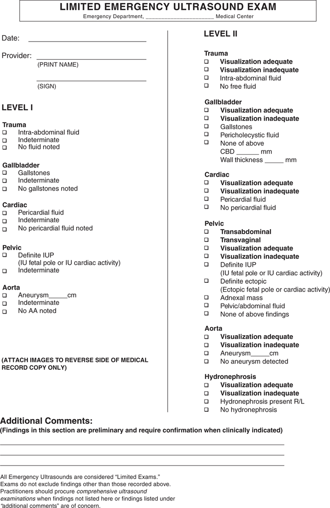
Figure 1-1. Example of bedside ultrasound report form.
Several options exist for the saving of images, including thermal printing and myriad of digital recording options. If results are to be included in the medical record, thermal images provide the easiest option, although with the growing use of electronic medical records, it may be possible to incorporate digital images into the medical record. For some applications, images need to be posted on the medical record in order to bill.9 If images are to be archived separately from the medical record, digital storage or videotaping can be used; however, archiving images outside the medical record creates compliance problems with medical record confidentiality and lack of access by other clinicians. Some programs use digital video recording for quality assurance and teaching purposes only.
QUALITY IMPROVEMENT PLAN
Any department implementing a new ultrasound program should place a strong emphasis on QI. No other area of emergency ultrasound training will provide as many teaching opportunities as a well-run QI program. If the resources or experience are lacking initially to overread all of the program’s ultrasound images, then finding a trained colleague in another location to assist with this endeavor is an option. There are pitfalls common to every application of ultrasound that can serve as a springboard for providing feedback and teaching within the program. This is clearly one of the most advantageous methods of enhancing both the technical and interpretative skills of clinicians.
The cornerstone of the QI program is review of examinations by the program’s director. For a very active group of clinicians performing focused ultrasound examinations, it will be logistically difficult to review every ultrasound examination so a method for selecting examinations for review must be decided upon. Examinations can be reviewed on either an indicated basis or a random basis. Indicated reviews occur when a certain indicator is met, such as a reported discrepancy between the focused ultrasound examination and another definitive study or procedure, or when a case is referred by a colleague because of questions regarding the accuracy of the examination. Random reviews are conducted by randomly selecting a predetermined number, or percentage, of examinations to assess the overall performance of the group.
Problems that are encountered during the QI process should be categorized as to their importance. The following represents one method of categorization.
Level I: Minor
Level I deviations usually consist of some problem with the technical component of the examination (e.g., gain too high) or disagreement on diagnostic criteria (e.g., labeling a common bile duct as mildly dilated at 6.5 mm when for the patient’s age, this was a normal measurement). Level I problems have no direct bearing upon the medical management of the patient. Typically, when this level of disagreement is found, a documented written or electronic copy of the disagreement is sent to the recipient.
Level II: Moderate
Level II deviations consist of discrepancies in interpretation between the clinician’s recorded image(s) and the QI review. In these cases, the undiagnosed or misdiagnosed pathology is nonemergent. For example, the clinician may record a gallbladder examination in which gallstones were diagnosed. Upon review, the QI review discovers a classic novice pitfall of a hyperechoic duodenal area that is mistaken for gallstones. Typically, when this level of disagreement is found, a chart review is undertaken to determine if subsequent care of the patient was appropriate. Depending on the follow-up that was provided and the patient’s clinical condition at the time of disposition, the action taken can range considerably.
Level III: Major
Related posts:
Stay updated, free articles. Join our Telegram channel

Full access? Get Clinical Tree


