1. Measurement of bladder volume
2. Measurement of post-void residual
3. Measurement of prostate size and morphology
4. Assessment of anatomic changes associated with bladder outlet obstruction
(a) Bladder wall thickness
(b) Bladder wall trabeculation
(c) Bladder wall diverticula
(d) Bladder stones
5. Documentation of efflux of urine from the ureteral orifices
6. Evaluation of pediatric posterior urethral valves
7. Assessment of correct position of a urethral catheter
8. Guidance for placement of a suprapubic tube
9. Assessment for completeness of the evacuation of bladder clots
10. Evaluation of hematuria
11. Evaluation for bladder tumors
12. Evaluation for distal ureteral dilation
13. Evaluation for foreign body in bladder
14. Evaluation for distal ureteral stone
15. Evaluation for ureterocele
16. Evaluation for complete bladder emptying
17. Assessment for bladder neck hypermobility in women
18. Evaluation of pelvic fluid collections
19. Guidance for transperineal prostate biopsy
20. Imaging of prostate when the rectum is absent or obstructed
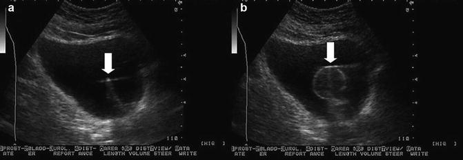
Fig. 8.1
(a) The tip of the balloon catheter is seen in the bladder (arrow). (b) Image of the inflated balloon in the bladder (arrow)
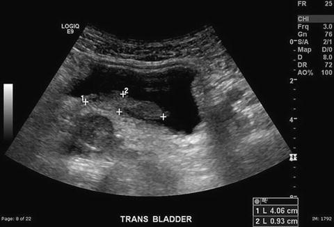
Fig. 8.2
Residual blood clot in the bladder after irrigation for clot retention
Pelvic ultrasound may also be useful to guide procedures such as deflation of a retained catheter balloon or for guiding the placement of a suprapubic catheter [1].
Patient Preparation and Positioning
The patient should have a full bladder but should not be uncomfortably distended. A bladder volume of approximately 150 cc is optimal. The patient is placed in the supine position on the examining table. The abdomen is exposed from the xiphoid process to just below the pubic bone. The patient may place their arms up above their head or by their side on the table. The room should be at a comfortable temperature. The lights are dimmed. A paper drape placed over the pelvic area and tucked into the patient’s clothing will protect the clothing and allow for easy cleanup after the procedure. The examiner assumes a comfortable position to the patient’s right.
Equipment and Techniques
The appropriate mode for performing pelvic ultrasound is selected on the ultrasound equipment, and the patient’s demographics are entered into demographic fields. A curved-array transducer is utilized for the pelvic ultrasound study (Fig. 8.3). The advantage of the curved-array transducer is that it requires a small skin surface for contact and produces a wider field of view. In the adult patient, a 3.5–5.0 MHz transducer is utilized to examine the bladder. For the pediatric patient, particularly for a small, young child, a higher frequency transducer is desirable, particularly for a small, young child.
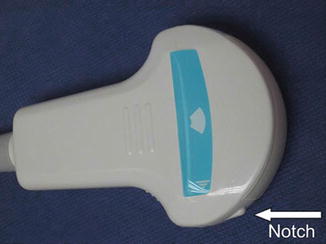

Fig. 8.3
Curved-array transducer with orienting notch (arrow). The notch is, by convention, oriented to the patient’s head during longitudinal scanning and to the patient’s right during transverse scanning
An even coating of warm conducting gel is placed on the skin of the lower abdominal wall or on the probe face. Prior to beginning the ultrasound examination the transducer is most often held in the examiner’s right hand. The orienting notch on the transducer is identified (Fig. 8.3). The orientation of the transducer may be confirmed by placing a finger on the contact surface near the notch. The image produced by contact with the finger should appear on the left side of the screen indicating the patient’s right side in the transverse plane and the cephalad direction in the sagittal plane.
Various techniques for probe manipulation are useful in ultrasound (Fig. 8.4). The techniques of rocking and fanning are non-translational, meaning the probe face stays in place while the probe body is rocked or fanned to evaluate the area of interest. The techniques of painting and skiing are translational, meaning the probe face is moved along the surface of the skin to evaluate the area of interest. All four techniques are useful for avoiding obstacles like the pubic bone or bowel gas and for performing a survey scan.
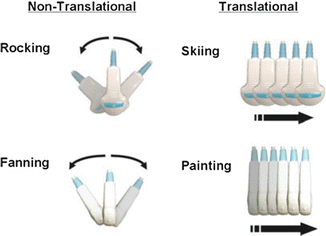

Fig. 8.4
Various techniques for probe manipulation
Ultrasound of the bladder may be started in the transverse view with the notch on the transducer to the patient’s right (Fig. 8.5). The transducer is placed on the lower abdominal wall with secure but gentle pressure. If the pubic bone is in the field of view, the bladder may not be fully visualized. The pubic bone will reflect the sound waves resulting in acoustic shadowing which obscures the bladder. The pubic bone may be avoided by varying the angle of insonation using the fanning technique until a full transverse image is obtained. In the transverse view of the bladder, the right side of the bladder should appear on the left side of the screen. The machine settings may be adjusted until the best-quality image is obtained.
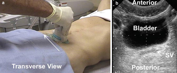

Fig. 8.5
(a) Position of transducer for obtaining a transverse image. Notch (arrow) is directed toward patient’s right. (b) Transverse image of the bladder with measurements of the width (1) and height (2) of the bladder. SV seminal vesicles
Survey Scan of the Bladder
A survey scan should be conducted prior to addressing specific clinical conditions to determine if any additional or incidental pathology is present. The survey scan is performed in the transverse view then the sagittal view. When starting with a transverse view, the sagittal view is obtained by rotating the transducer 90° clockwise with the probe notch pointing toward the patient’s head (Fig. 8.6). The rocking technique may be used to facilitate viewing the bladder in the sagittal view.
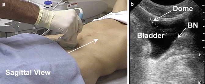

Fig. 8.6
(a) Position of the transducer with the notch to the patient’s head. (b) Sagittal image of the bladder with length of the bladder (3) from the dome on the left to the bladder neck (BN) at the right
Measurement of Bladder Volume
The bladder volume is obtained by first locating the largest transverse diameter in the mid-transverse view (Fig. 8.7). The width and the height are measured. The transducer is then rotated 90° clockwise to obtain the sagittal view. In the midsagittal view, the length of the bladder is measured using the dome and the bladder neck as the landmarks. These measurements may be made using a split screen so that both measurements are on the same screen. These images are printed or saved electronically. The bladder volume is calculated by multiplying the width, height, and length measurements by 0.625. When the specified measurements are obtained, the calculated bladder volume will usually be automatically displayed. When measuring urine volume in the bladder, the report should indicate whether the volume is a bladder volume measurement or a post-void residual urine volume measurement.
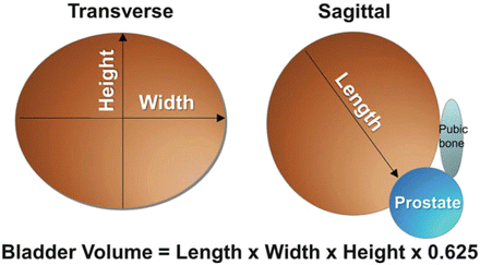

Fig. 8.7
The formula for calculating bladder volume = width (1) × height (2) × length (3) × 0.625
Measurement of Bladder Wall Thickness
Measurement of bladder wall thickness is taken, by convention, when the bladder is filled to at least 150 cc. Bladder wall thickness may be measured at a number of different locations. In this case, the bladder wall thickness is measured along the posterior wall on the sagittal view (Fig. 8.8). If the bladder wall thickness is less than 5 mm when the bladder is filled to 150 cc, there is a 63 % probability that the bladder is not obstructed. However, if the bladder wall thickness is over 5 mm at this volume, there is an 87 % probability that there is bladder outlet obstruction. Nomograms are available to calculate the likelihood of urodynamically demonstrated bladder outlet for bladder wall thickness at various bladder volumes [2].
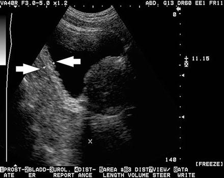

Fig. 8.8


Bladder wall thickness , in this case, is measured (arrows) along the posterior wall. In this image the wall measures 11.15 mm in thickness
Stay updated, free articles. Join our Telegram channel

Full access? Get Clinical Tree


