Transient Ischemic Attack and Perfusion Imaging
Shyam Prabhakaran
Jonathan Kleinman
Greg Zaharchuk
Jean-Marc Olivot
Transient ischemic attack (TIA) has been traditionally defined as focal neurologic symptoms attributed to a cerebrovascular territory lasting less than 24 hours.1,2 In its recent review, the American Heart Association/American Stroke Association reports that for TIAs lasting typically minutes the presence of an infarction on magnetic resonance imaging (MRI) is not correlated with symptom duration.3 These experts redefined TIA by focal brain, spinal cord, or retinal ischemia without evidence of tissue infarction on brain imaging.3 Nevertheless, as most of the epidemiologic studies refer to it, this chapter will use the classical definition of TIA.
As brain infarction and TIA form a continuum, their mechanisms and risk factors are similar. Epidemiologic studies estimate that approximately 200,000 TIA patients present annually in the United States.4,5 Another study from the National Hospital Ambulatory Medical Care Survey documented over 2.5 million TIAs and calculated an overall incidence rate of 1.1 per 1000 US population.6 In contrast to the nearly 30% reduction in the incidence of first-ever stroke, the incidence of TIA appears to be increasing based on a population based-study in the United Kingdom.7
In most TIAs, symptoms have resolved and physical examination is normal at the time of the clinical evaluation. Thus, the diagnosis of TIA is based on the patient’s recollection and description of symptoms. Some features, including type of symptoms (motor and language), sudden onset and short duration of symptoms, and associated vascular risk factors, can help in the clinical diagnosis.8 Up to 60% of patients referred to neurologists for evaluation of TIA have alternate diagnoses.9,10,11,12 Therefore, agreement on a TIA clinical diagnosis remains poor, even among stroke neurologists, whose judgments are often considered the gold standard.13,14 With the advent of diffusion-weighted imaging (DWI) and computed tomography (CT) or MR perfusion imaging, the finding of cytotoxic injury (restricted diffusion) or focal hypoperfusion in patients evaluated for suspicion of TIA provides convincing evidence of brain ischemia.15,16,17,18,19,20 Therefore, the combination of a specialized clinical evaluation with advanced neuroimaging is the mainstay of TIA evaluation.3
TIA diagnosis is an emergency because the risk of stroke after a TIA is high (20% at 3 months)21 and approximately half of this risk occurs within the first 48 hours of TIA symptoms.22,23,24 Several tools exist to estimate the risk of stroke after TIA. The ABCD2 (Age, Blood pressure, Clinical symptom type, Duration of symptoms, and Diabetes) score is a simple clinical scale that evaluates the characteristics of a TIA. In the referent publication, the risk of stroke at 2 days associated with ABCD2 score was, respectively, for a score of six or seven, 8.1%, four or five, 4.1%, and zero to three, 1.0%. Therefore, an ABCD2 score above three has been chosen to decide patient hospitalization.15 The ABCD2 score has become the standard tool to estimate the risk of stroke after a TIA.25 Nevertheless, this score accurately predicts stroke in only 60% of patients and, therefore, may not be considered by itself a reliable tool to triage TIA patients.26,27,28 The detection of tissue infarction or symptomatic brain or neck vessel stenosis represents independent risk factors of stroke.16,19,26,29,30,31,32 The incorporation of brain infarction and symptomatic vessel stenosis into the ABCD2 score increases its ability to predict the risk of future stroke by almost 15%.31,32
Magnetic Resonance Imaging
Multimodal MRI is both more specific and sensitive than head CT for the detection of acute ischemia.33 DWI reveals an acute infarction in about 30% of the patients with a discharge diagnosis of TIA.30 One recent meta-analysis showed that 7% of TIA patients with a positive DWI will suffer a stroke within a week versus only 0.5% of the DWI-negative patients.31 Interestingly, the ABCD2 score does not influence the risk of stroke in DWI-negative patients, suggesting that a proportion of these patients may not have experienced a transient ischemic event.30 Based on its sensitivity to detect brain infarction in TIA patients, DWI combined with urgent vessel imaging (cervical and intracranial) is recommended for the emergent evaluation of every TIA patient.3
Perfusion Imaging
As mentioned throughout this book, perfusion-weighted imaging (PWI) gives an estimate of cerebral hemodynamics and can be abnormal in several situations.
Acute Ischemia
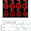
In the absence of a vessel occlusion on MR angiography (MRA) or DWI lesion, perfusion imaging can reveal persistent ischemia that may reflect a distal occlusion not captured by the MRA or the absence reperfusion after recanalization of the occluded vessel. This no-reflow phenomenon has been described in coronary disease after
successful recanalization of the occluded vessel and refers to the persistent occlusion of the capillary vessels due to inflammatory and thrombotic mechanisms.34 In this situation, perfusion imaging may help the physician to confirm the diagnosis of ischemia (Fig. 50.1).35
successful recanalization of the occluded vessel and refers to the persistent occlusion of the capillary vessels due to inflammatory and thrombotic mechanisms.34 In this situation, perfusion imaging may help the physician to confirm the diagnosis of ischemia (Fig. 50.1).35
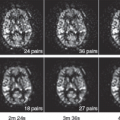
The mismatch between DWI and PWI outlines the extent of the tissue at risk for infarction. This region, called the penumbra, may progress to infarction in the absence of reperfusion. In this situation, perfusion imaging can help to predict the risk of future infarction (Fig. 50.2).36,37
Stay updated, free articles. Join our Telegram channel

Full access? Get Clinical Tree


