(1)
Department of Radiology, John H Stroger Jr Hospital of Cook County, Chicago, IL, USA
Liver
Diagnosis
Penetrating Grade III Liver Laceration
Imaging Features
1.
Hematoma: subcapsular >50 % surface area and rupture with active bleeding
2.
Hematoma: intraparenchymal >10 cm
3.
Laceration: capsular tear, >3 cm in depth
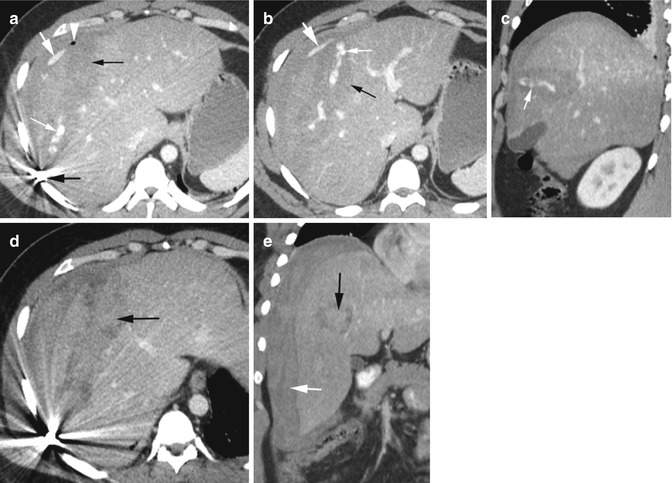
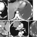
Fig. 11.1
Penetrating liver laceration grade III by gunshot injury. Contrast-enhanced CT (a, b) axial and (c) coronal reformatted images show linear low-density laceration of the liver (thin black arrow) with high-density contrast mixed with active hemorrhage (thin white arrow) within it which is also extending into the subphrenic space (thick white arrow), a bullet adjacent to the rib (thick black arrow) and with small focus of gas (arrowhead). (d, e) Axial and coronal reformatted CTs 1 week later show high-density hematoma in the laceration (black arrow) and perihepatic space (white arrow) but no active hemorrhage
Diagnosis
Laceration of the Liver, Tear of the Diaphragm, and Pulmonary Contusion
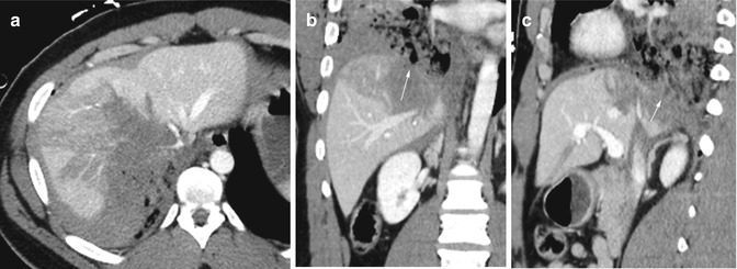
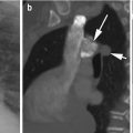
Fig. 11.2
Liver laceration, lung contusion, and tear of the diaphragm from penetrating trauma. (a) Axial CT and (b) coronal and (c) sagittal reformatted images show low-density laceration of the dome of the right lobe of the liver continuous with the adjacent pulmonary contusion. The diaphragm is discontinuous posteriorly (arrow)
Diagnosis
Subcapsular Hemorrhage
Imaging Features
1.
High-density hemorrhagic fluid extending from the liver parenchyma into the subcapsular space
2.
Fluid contouring the liver and deforming the liver surface
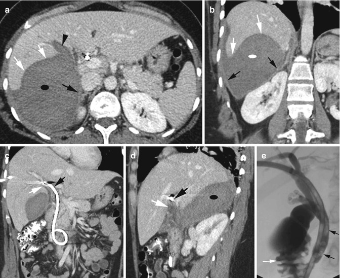
Fig. 11.3
Subcapsular hemorrhage from PTC drain placement. History of laparoscopic cholecystectomy and impacted stone in CBD followed by PTC drain placement. (a, b) Axial CT and coronal reformatted images show a thin linear capsule (black arrows) surrounding the hemorrhagic fluid (ellipse also in d) that is compressing and deforming the liver surface (white arrows) and is extending from the liver parenchyma (arrowhead). (c, d) Coronal and sagittal reformatted images show hemorrhage tract (white arrow) extending from near the biliary drain (black arrow). (e) T-tube cholangiogram shows multiple stones in the common bile duct as filling defects (black arrows) with contrast flowing into the duodenum (white arrow)
Diagnosis
Biloma
Imaging Features
1.
Laceration of the liver parenchyma
2.
Large well-marginated fluid collection at the site of the previous laceration
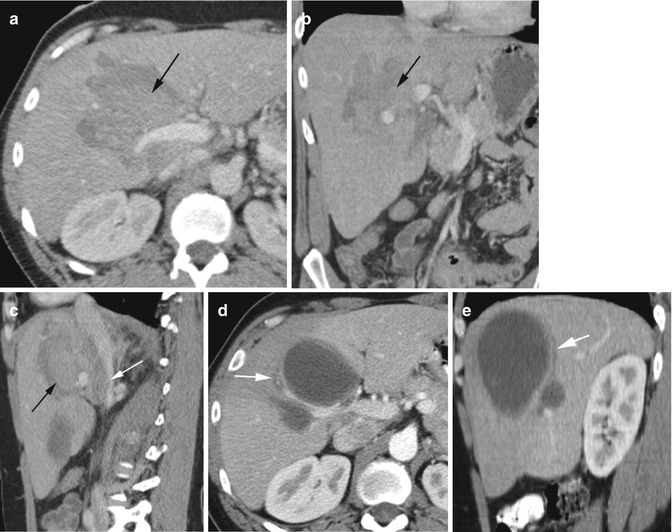
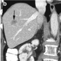
Fig. 11.4
Liver laceration grade IV with high-density hematoma from blunt trauma and well-marginated biloma replacing the hematoma a month later, which was drained. Axial CT and coronal and sagittal reformatted images (a– c) show laceration with hematoma (black arrow) involving porta hepatis and causing mass effect on the IVC (white arrow in c). (d, e) Axial and sagittal reformatted images 1 month later show two loculated low-density fluid collections continuous with the biliary duct (white arrow) which were drained
Diagnosis
Injury to the Biliary Tract
Imaging Findings
1.
Air in the biliary tree
2.
Biliary fluid and gas contained within the liver capsule
3.
Laceration of the liver from penetrating trauma
4.
Spillage of contrast from the bile ducts by ERCP study
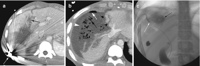
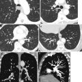
Fig. 11.5
Laceration of the liver and injury to the biliary ducts. Axial CT (a) shows laceration of the right lobe of the liver (black arrow) by a bullet (white arrow). (b) At a higher level than (a), gas is seen in the biliary tree (black arrow), and large biloma is seen with layered gas (white arrow) distending the liver capsule and outside the liver parenchyma (arrowhead). (c) ERCP examination shows leakage of contrast (black arrow) with drainage catheter (white arrow)
Diagnosis
Subcapsular Hepatic Active Bleeding by Angiogram
Imaging Features
1.
Loculated parenchymal and subcapsular hematoma
2.
No active hemorrhage in postcontrast study
3.
Active hemorrhage by angiogram
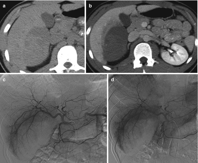
Fig. 11.6
Hematoma involving the liver parenchyma and subcapsular region with active bleeding by angiogram, with history of blunt trauma. Axial CT (a) precontrast shows high-density hematoma involving segment VI and protruding inferiorly with subcapsular extension. (b) Postcontrast study does not show active hemorrhage within the hematoma. (c) Angiogram of the hepatic artery shows faint blush of extravasation in the early part of the study. (d) Delayed phase of the study shows increased extravasation of contrast
Spleen
Diagnosis
Splenic Laceration
Imaging Features
1.
Laceration of the spleen with or without active intraparenchymal bleed from disruption of trabecular vessels
2.
Capsular tear with active intraperitoneal bleed
3.
Parenchymal and subcapsular hematoma

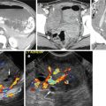
Fig. 11.7
Splenic laceration in different patients. Patient 1: (a) axial contrast-enhanced CT following blunt trauma shows grade III laceration with injury to the trabecular vessels and intraparenchymal pooling of contrast (black arrows). Cortical tear is also seen with active bleeding into the peritoneal cavity (white arrow). Patient 2: (b, c) axial and (d) coronal reformatted CTs following penetrating wound show active bleeding from the spleen into the peritoneal cavity (arrows). Patient 3: (e) axial noncontrast image shows linear high-density hematoma within laceration (arrow). (f, g) CECT shows linear tear of the spleen (arrow) and large subcapsular hemorrhage (ellipse)
Pancreas
Diagnosis
Pancreatic Tear
Imaging Features
1.
Linear lucent line through the neck with complete separation of the neck and body
2.
Laceration of the pancreas with several incomplete lines through the pancreas
3.
Surrounding hemorrhage and/or pancreatic fluid
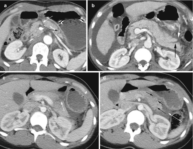
Fig. 11.8
Tear and laceration of the pancreas in three patients with trauma. Patient 1: (a) axial CT shows complete tear of the neck (white arrow) and at the tail (black arrow). The surrounding low-density fluid is leaking pancreatic fluid from the tear of the pancreatic duct. Patient 2: (b) axial CT following a stab wound shows incomplete tear at the tail of the pancreas (black arrow) with surrounding small high-density hematoma (white arrow). Patient 3: (c, d) axial CT shows multiple incomplete tears of the tail of the pancreas (black arrows) with surrounding high-density hematoma (white arrow)
Kidneys
Diagnosis
Renal Laceration and Hematoma
Imaging Features
1.


Sharp tear through the renal parenchyma
Related posts:
Stay updated, free articles. Join our Telegram channel
Full access? Get Clinical Tree
