High-energy injuries: Motor vehicle accidents constitute ∼ 50% of cases, with seat belts increasing risk
– “High-riding” seat belt incorrectly placed over abdomen increases risk (muscle avulsion from iliac crest)
– Other traumatic injuries are common (∼ 80%), with up to 50% of patients suffering other abdominal injuries requiring surgery
Low-energy injuries (most common in children): Impact by small blunt object (such as bicycle handlebar, i.e., handlebar hernia)
– Hernias can develop after minor trauma in children
CLINICAL ISSUES
• May be overlooked clinically at time of injury and often diagnosed due to hernia-related complications
Only 22% of patients in 1 series had TAWH diagnosed clinically, making CT essential to diagnosis
Complications: Incarceration; bowel strangulation, perforation, and ischemia
• Peak incidence in children < 10 years of age due to handlebar injuries
2nd most common age group is 20-50 years due to motor vehicle accidents
• Treatment: Delayed repair of hernia usually performed 6-8 weeks following high-energy injuries to allow primary tissue damage to subside
(Left) Axial CECT demonstrates small bowel and colon herniating through a traumatic abdominal wall defect. At surgery, several segments of small bowel had serosal tears and avulsions, requiring resection.
(Right) Axial CECT demonstrates a traumatic lumbar hernia, with herniated abdominal fat covered only by the latissimus dorsi muscle . Also noted is infiltration of the intraabdominal fat adjacent to the hernia. At surgery, a serosal tear of the descending colon was identified.
(Left) Axial CECT demonstrates a large amount of hypoenhancing small bowel herniated through a traumatic hernia of the right abdominal wall. Active arterial bleeding is evident. Much of the herniated bowel was not viable at the time of surgery.
(Right) Axial CECT shows disruption of the abdominal wall muscles in the left lower quadrant, with the muscles avulsed from their attachment to the iliac crest. Note the presence of adjacent subcutaneous hematoma . This is a typical example of a seat belt injury.
TERMINOLOGY
Abbreviations
• Traumatic abdominal wall hernia (TAWH)
Definitions
• Traumatic disruption of musculature and fascia of anterior abdominal wall due to blunt trauma (in absence of penetrating injury) ± herniation of bowel or visceral organs into subcutaneous space
Development of new abdominal wall hernia in patient with recent blunt trauma (without penetrating injury)
• Location
Roughly 75% occur in lower abdomen
– May reflect inherent weakness of lower abdomen due to natural orifices (such as inguinal canals) and susceptibility to increased intraabdominal pressures
Equally common in right and left sides of abdomen
Common locations include
– Region of iliac crest in seat belt injury (site of lap and shoulder strap junction)
– Focal hernias often occur in lower abdomen lateral to rectus sheath or inguinal region
– Larger, diffuse abdominal wall defects most often sustained in motor vehicle accidents
– Rarely, hernias may occur through tear in retroperitoneum
• Size
Anatomical defects vary from small defects (few centimeters) to large disruptions
Only gold members can continue reading. Log In or Register to continue
 Low-energy injuries (most common in children): Impact by small blunt object (such as bicycle handlebar, i.e., handlebar hernia)


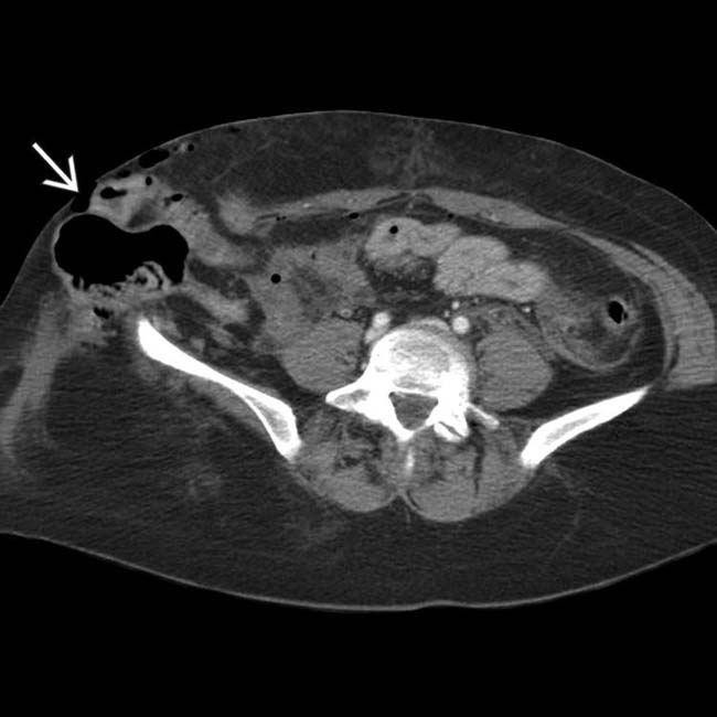
 herniating through a traumatic abdominal wall defect. At surgery, several segments of small bowel had serosal tears and avulsions, requiring resection.
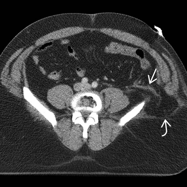
 . Also noted is infiltration of the intraabdominal fat
  adjacent to the hernia. At surgery, a serosal tear of the descending colon was identified.
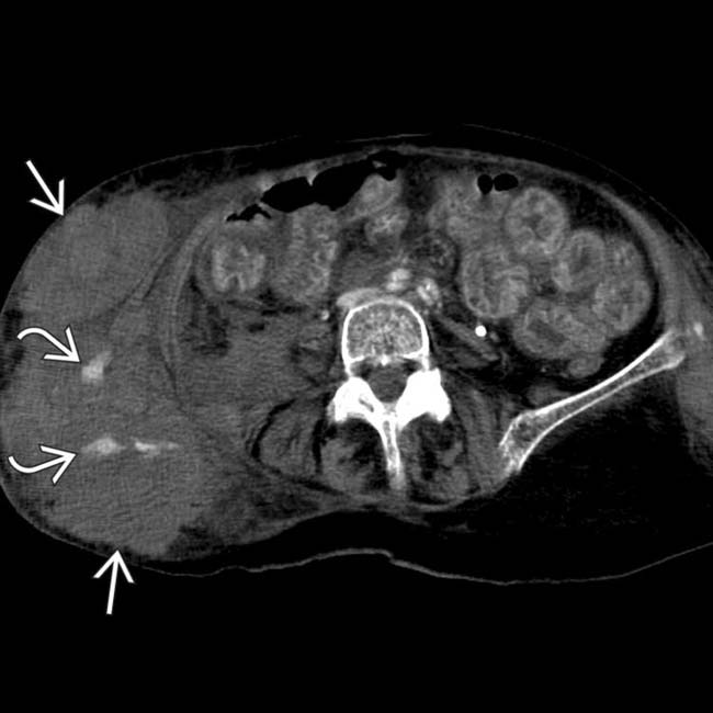
 herniated through a traumatic hernia of the right abdominal wall. Active arterial bleeding
  is evident. Much of the herniated bowel was not viable at the time of surgery.
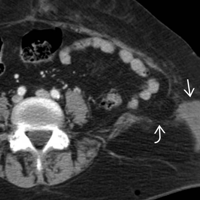
 in the left lower quadrant, with the muscles avulsed from their attachment to the iliac crest. Note the presence of adjacent subcutaneous hematoma
  . This is a typical example of a seat belt injury.


