DSA demonstration of AVF nidus with enlarged draining veins
Unlike spontaneous DAVF (sDAVF), often no cord enlargement/hyperintensity from congestive myelopathy
• Angiographic protocol for evaluation of suspected cervical tDAVF includes selective injections of both external carotid + vertebral arteries, thyrocervical trunks, and paired segmental arteries
Angiographic appearance is similar to sDAVF, but nidus may be extraforaminal/paravertebral instead of along nerve root
Pathology
• Any segment of spine, most frequently cervical
• Usually caused by direct penetrating wounds
• May also occur with blunt trauma and vertebral fracture
Clinical Issues
• Symptoms may relate to
Venous hypertensive myelopathy
Compression by enlarged draining veins
Steal phenomenon
• Endovascular treatment with embolization or surgical ligation
(Left) Off-midline sagittal T1WI MR shows an enlarged ventral epidural venous plexus with a prominent flow void in this patient with a C2 fracture.
(Right) Axial T2* GRE MR shows dilation of the epidural venous plexus with prominent flow voids causing compression of the nerve root in the lateral recess of the cervical spine.
(Left) Anteroposterior DSA from the left vertebral artery shows high-flow arteriovenous communication near the V2/V3 junction of the right vertebral artery , with reversed flow in the distal right vertebral artery supplying dilated epidural plexus and dilated deep cervical veins.
(Right) Coronal CT myelography of the lumbar spine shows dilated, tortuous veins in the caudal thecal sac due to a post-traumatic (iatrogenic) arteriovenous fistula.
TERMINOLOGY
Abbreviations
• Arteriovenous fistula (AVF)
• Traumatic dural AV fistula (tDAVF)
Synonyms
• Vertebrovertebral arteriovenous fistula
Definitions
• Trauma-induced arteriovenous fistula
IMAGING
General Features
• Best diagnostic clue
DSA demonstration of AVF nidus with enlarged draining veins
• Location
Dural or extradural nidus
Any segment of spine, most frequently cervical
• Size
Small nidus
May have extensive draining venous engorgement
• Morphology
Small, tight AVF nidus
Only gold members can continue reading. Log In or Register to continue
 Unlike spontaneous DAVF (sDAVF), often no cord enlargement/hyperintensity from congestive myelopathy


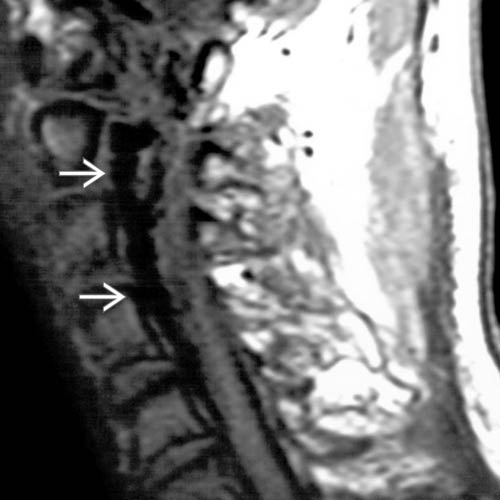
 in this patient with a C2 fracture.
 causing compression of the nerve root in the lateral recess of the cervical spine.
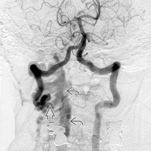
 , with reversed flow in the distal right vertebral artery supplying dilated epidural plexus
  and dilated deep cervical veins.
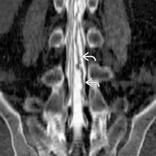
 due to a post-traumatic (iatrogenic) arteriovenous fistula.