, Valery Kornienko2 and Igor Pronin2
(1)
N.N. Blockhin Russian Cancer Research Center, Moscow, Russia
(2)
N.N. Burdenko National Scientific and Practical Center for Neurosurgery, Moscow, Russia
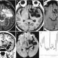
Acute hemorrhagic lesions from multiple contusions (up to 3 days) of the brain can be visualized by MRI with a certain degree of similarity of the tissue characteristics to those in metastatic lesions. A situation, when patients can be transported to the hospital in an unconscious state, cannot be excluded, since this can also occur as a result of strokes and hemorrhages into tumor lesions.
Such lesions in the acute stage contain mostly intracellular paramagnetic deoxyhemoglobin, which is formed upon dissociation of oxygen and hemoglobin. Since deoxyhemoglobin in undamaged hypoxic red cells does not cause a shortening of T1, hemorrhagic injuries have a normal or weakly hypointense signal in this sequence. The high concentration of red blood cells and fibrin in the clot is causing shortening of T2 and the emergence of very low signal areas in T2-weighted and T2*-weighted, T2-FLAIR images (Fig. 42.1). A few days later, the lesion of subacute contusion begins to “liquefy” with the development of a vasogenic edema that increases within the first week, which may be the cause of cerebral herniations. With the transformation of deoxyhemoglobin to the paramagnetic intracellular methemoglobin, the interaction between hydrogen atoms and the paramagnetic methemoglobin center is the cause of high signal intensity in T1-weighted images, which first manifests along the periphery of the contusion. Intracellular methemoglobin is characterized by a low signal in T2-weighted images.