104 Truncation Artifacts
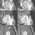
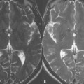
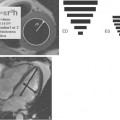
Figure 104.1A suffers from prominent truncation artifacts (black arrows) in the form of ringing (or “ring-down”), seen as alternating light and dark bands within the periphery of the brain. Figure 104.2A illustrates a syrinx-like artifact (white arrows) within the center of the thoracic cord on a T1-weighted sagittal scan, a classic truncation artifact that appears as a result of alternating dark and light signal bands overlying the spinal cord. This artifact is easily mistaken for hydromyelia (dilatation of the central canal, more commonly but incorrectly referred to as a “syrinx”), with the scan obtained in this instance in a normal, healthy volunteer. The artifacts in both scans result from an inappropriate choice of pixel size (too large). Truncation artifacts are reduced by the use of a smaller field of view (FOV) or a larger matrix size, the latter approach illustrated in both Fig. 104.1B
Stay updated, free articles. Join our Telegram channel

Full access? Get Clinical Tree


