, Valery Kornienko2 and Igor Pronin2
(1)
N.N. Blockhin Russian Cancer Research Center, Moscow, Russia
(2)
N.N. Burdenko National Scientific and Practical Center for Neurosurgery, Moscow, Russia
Metastases to the pineal region of malignant tumors are quite rare, occurring not more than in 1%. Primary tumors of the pineal region are also relatively rare, accounting for 0.5–1% of all intracranial tumors; in children, their proportion in the structure (3–8%) is slightly higher. Both benign and malignant (75%) tumors develop in the pineal region (Jooma et al. 1983; Bruce et al. 1993; Allen et al. 1996; Konovalov and Pitskhelauri 2004). According to the modern histological classification (2016), tumors of the pineal region are presented by four major groups: (a) pineocytoma, (b) parenchymal tumor, (c) pineoblastoma, and (d) papillary tumor. Four main syndromes prevail in the clinical picture of the pineal region lesions: (a) intracranial hypertension due to compression of the cerebral aqueduct and hydrocephalus, (b) Parinaud syndrome (midbrain involvement), (c) cerebellar disorders, and (d) endocrine disorders (premature sexual development, diabetes insipidus).
36.1 Glial Tumors
Glial tumors spread to the pineal region from the neighboring structures and brain structures—the midbrain, thalamus, optic thalamus, quadrigeminal plate, and splenium of the corpus callosum. This series of tumors are mainly represented by astrocytomas and ependymomas. As a rule, in the group of astrocytic gliomas of the pineal region, benign forms are more common—diffusely growing gliomas of the optic thalamus or quadrigeminal plate; malignant ones (glioblastoma ) are much rarer. CT and MRI characteristics of malignant astrocytomas, including glioblastoma, are characterized by various structures and types of accumulation of the contrast agent (Fig. 36.1).
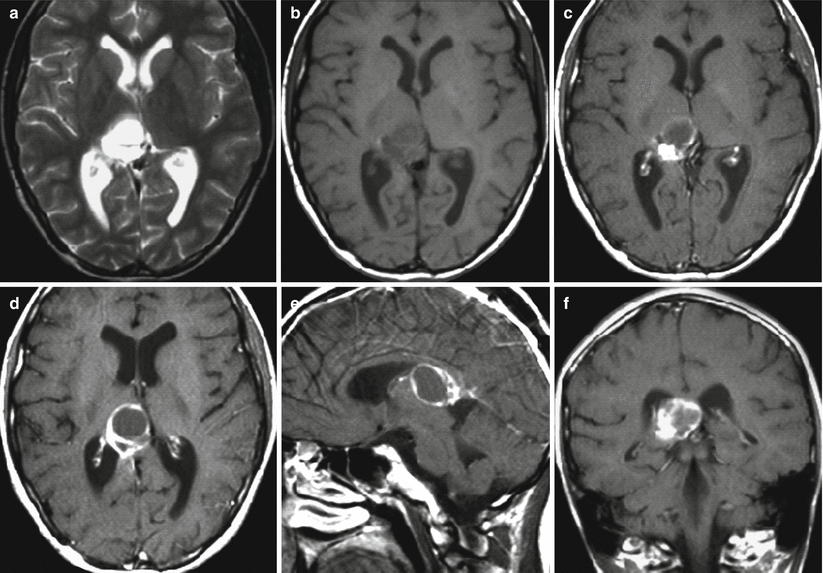

Fig. 36.1
Glioblastoma. On T2-weighted MRI (a) and T1-weighted MRI before (b) and after (c–f) intravenous contrast enhancement, in the projection of the right optic thalamus, there is a space-occupying lesion with a necrotic center and an intensely contrast-enhanced infiltrative peripheral part. There is no perifocal edema. The posterior sections of the third ventricle and quadrigeminal plate are deformed
36.2 Germ-Cell Tumors
Germ-cell tumors are dysembryogenetic tumors that mostly affect the reproductive organs but can also be identified within the CNS, while being usually located medially. In the central nervous system, they are more likely to occur in the pineal region, followed by the suprasellar region, and they are rarely diagnosed in the optic thalami and basal ganglia, ventricular system, cerebellum, etc. According to their histological structure, germ-cell tumors can be divided into several subtypes: germinoma, embryonal carcinoma, endodermal sinus tumor, choriocarcinoma, teratoma, and mixed tumors. Embryonal carcinoma, endodermal sinus tumor, and choriocarcinoma are regarded as non-germinal germ-cell tumors.
36.2.1 Germinomas
Germinomas are observed in up to 67% of cases among germ-cell tumors, and their typical site is the pineal region; thus, they make up about 40% of all tumors in this area, though they may also be observed in other brain regions: suprasellarly—from 25% to 35%, in the projection of the basal ganglia—up to 10% of cases. Germinomas are non-encapsulated lesions, grow usually expansively, but may have infiltrative growth. They often become calcified and have hemorrhages. Histologically, testicular germinomas are identical to seminomas and consist of cords or meshes of glycogen-containing large tumor cells with large vesicular nuclei, surrounded by a fibrous connective tissue with lymphocyte accumulations located in-between (Macko 1998).
Stay updated, free articles. Join our Telegram channel

Full access? Get Clinical Tree


