Fig. 24.1
Abdominal organs (ileum, spleen, and cholecyst) and lymphatic node lesions of typhoid fever. (a–g) Ultrasound demonstrates thicker wall of the ileum, enlarged mesenteric lymphatic nodes, enlarged spleen with normal echoic structure, enlarged cholecyst, thicker wall of the cholecyst with more blood vessels in it, cholestasis, and ulcers in cholecystic mucosa with interruption of its continuity (Reprint with permission from Mateen MA, et al. Indian J Pediatr, 2006, 73 (8): 681)
CT Scanning
CT scanning demonstrates enlarged lymphatic nodes and annular thickening of the intestinal wall, especially at the terminal ileum; diffuse enlargement of the spleen with uneven density of some nidi and sheet nidi with low density that reveal splenic abscess or destruction, which can be identified by enhancement scanning; moderate enlargement of the liver compared with enlarged spleen; and even thickening of the cholecystic wall with even low density by enhancement scanning. Some results show ascites and a few reveal intestinal perforation, bleeding, and abscess (Figs. 24.2, 24.3, and 24.4).
Case Study 2
A 29-year-old male with fever and right lower quadrant pain.
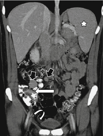
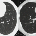
Fig. 24.2
Abdominal organs (ileum, spleen, and cholecyst) and lymphatic node lesions of typhoid fever. CT scanning of the abdomen demonstrates thickening of the distal ileum wall (white arrows), enlarged mesenteric lymphatic nodes (black arrows), enlarged spleen (stars), and effusions (white curving arrows)
Case Study 3
A 22-year-old male with clinical diagnosis of typhoid fever.
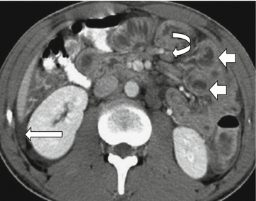
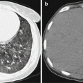
Fig. 24.3
Typhoid fever involving jejunum and lymph nodes. CT scanning demonstrates thickening of the jejunum wall (short arrows), enlarged lymphatic nodes (curving arrow), and ascites (long arrow)
Case Study 4
A 43-year-old male with abdominal distension.
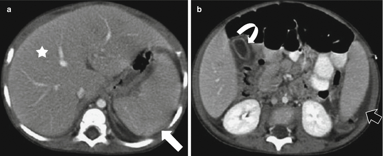
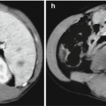
Fig. 24.4
Abdominal organs (ileum, spleen, and cholecyst) and lesions of typhoid fever. (a, b) CT scanning demonstrates enlarged liver (star) and spleen (white arrow), thickening of the cholecyst wall (curving arrow), and ascites (black arrow)
24.7.1.2 Typhoid Encephalopathy
Examinations reveal mild hydrocephalus, slightly expanded encephalocele but without sulcus and cistern expansion, localized and diffuse decrease of white matter density, and parts of enhanced nidi. In addition, enhanced meninx line and diffusely enhanced gyri are often seen, while basilar cistern and ependyma are rarely enhanced. CT scanning reveals common features of encephalitis, meningitis, and meningoencephalitis, which may exist together or solely.
24.7.1.3 Pulmonary Infection
Pulmonary infections often involve pulmonary mesenchyme, which is characterized by bronchial vascular bundles and interlobular and intralobular septum thickening. Reticular opacity and reticular nodes with clear edges and slightly high density can be seen in the lungs.
Case Study 5
A patient with typhoid fever with pulmonary infections.
For case detail and figures, please refer to Zou Jun. Infection and Chemotherapy Journal of China. 2008, 8 (3): 228.
24.7.2 Paratyphoid Fever
Imaging demonstrations of paratyphoid fever A, B, and some paratyphoid fever C are similar, which are generally characterized by annular thickening of the intestinal wall, especially the terminal ileum. Mesenteric lymphatic nodes, spleen, and liver enlarge. Some of the cases are present with ascites and a few with intestinal perforation, bleeding, and abscess. Septicopyemia caused by paratyphoid fever C can mainly induce suppurative inflammation, which is characterized by systemic suppurative metastatic nidi, of which localized suppurative inflammation in the lungs, bones, and joints, meningitis as well as endocarditis are the most common (Fig. 24.5).
Related posts:

Stay updated, free articles. Join our Telegram channel

Full access? Get Clinical Tree


