14 Ultrasound-Guided Catheter Placement for Peripheral Nerve Blocks
Ultrasound-guided catheter placement is a complex procedure involving advanced manipulative skills of the operator.1 Common procedures are ultrasound-guided interscalene, femoral, and popliteal catheters.2 Both in-plane and out-of-plane approaches are popular for peripheral nerve catheter insertion.3
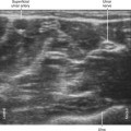
With in-plane technique the needle bevel can be turned so that the catheter slides along the nerve rather than around the nerve.4 A helically wound, metal-reinforced catheter can act as a spatially modulated wire.5 Therefore, the ultrasound beam will be reflected back to the transducer regardless of the catheter orientation to improve visibility.
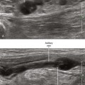
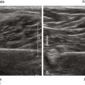
Long-axis in-plane approaches to peripheral nerve catheter placement have been reported.6 The advantage is that the peripheral nerve, block needle, and catheter can all be viewed at the same time. However, it can be difficult to manipulate the transducer to maintain all three within the plane of imaging. Furthermore, because all three are constrained to lie in the same plane, care must be taken not to advance the needle and catheter into the nerve. Sciatic nerve catheters are particularly amenable to the long-axis in-plane approach if the patient can be placed in a prone position. With this long-axis approach, the operator views directly across the patient for anatomic orientation of the ultrasound monitor.
Ultrasound guidance appears to have advantages over nerve stimulation for peripheral nerve catheter placement because the performance times are more consistent.7
Related posts:


Stay updated, free articles. Join our Telegram channel

Full access? Get Clinical Tree


