Introduction
Ultrasound machines with high-resolution probes are readily available in most radiology departments and are routinely used to assess both articular and periarticular disorders. They are also becoming commonplace in rheumatology departments, reflecting the important role they now play in rheumatological disease. Ultrasound has many advantages over other imaging techniques with the ability to carry out rapid assessment of multiple joints at different locations, undertake dynamic imaging and guide diagnostic and therapeutic injections. This chapter focuses on the role of ultrasound in joint disease and particularly rheumatological conditions, the technical aspects of joint ultrasound examination and the imaging findings.
Technical Aspects of Ultrasound
The development of higher-frequency transducers has allowed for improved resolution and, with the majority of joints lying relatively superficially, linear array probes of frequencies of 10 MHz or higher can be effectively utilized. Curvilinear probes, although rarely required for musculoskeletal imaging, may be useful for examining deeper joints such as the hip joint. Although transducer selection primarily depends upon the frequency, the probe footprint (the surface area of the transducer in contact with the skin) should be considered. Small footprint probes can be easily manoeuvred to image small superficial structures, small joints and at bony prominences such as the malleoli, where the skin surface does not allow adequate contact with larger probes. Stand-off gel pads can prove useful to reduce the amount of near-field reverberation when examining superficial structures; however, with modern probes these are rarely necessary and the use of liberal amounts of ultrasound jelly is usually all that may be required in practice.
Pitfalls and Limitations
Many of the pitfalls and limitations of ultrasound are dealt with elsewhere in this book. They include anisotropy and beam edge artifact. However, when undertaking ultrasound of joint disease certain specific pitfalls should be considered.
Excessive probe pressure can obliterate small quantities of fluid, reduce the sensitivity for detection of blood flow and may obscure synovitis.
| If it proves difficult to maintain probe contact with the skin surface using minimal probe pressure, a thick application of sonographic jelly may help. |
Expertise is important in the interpretation of both ultrasound and MRI; however, unlike with MRI, reevaluation of ultrasound requires the patient to be recalled. Thus standardization of ultrasound criteria and validation of training both of the radiologist and the rheumatologist who perform these studies are paramount.
Techniques for Scanning the Small Joints of the Hands and Feet
While an all-inclusive examination of the small joints may be desirable, this is daunting and time consuming and can be modified by omitting joints that are frequently uninvolved, such as the distal interphalangeal joints (IPJs) and thumbs in rheumatoid arthritis.
It is the authors’ routine to examine the index, middle, ring and little fingers, although this may be adapted for specific clinical indications. Superficial structures such as the tendon and tendon sheath are assessed prior to the joint itself, where standard sagittal images form the basis of the examination, with axial (metacarpophalangeal joint, MCPJ) and coronal and axial (proximal interphalangeal joint, PIPJ) images used as adjuncts.
| Routine examination of the extensor aspect of the MCPJs, followed by the extensor, ulnar and radial aspects of the IPJs, is performed. |
The radial aspect of the joint should be carefully scrutinized, as synovial hypertrophy and erosions are predominant at this site.
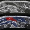
It is essential to appreciate the normal sonographic anatomy of the small joints to be able to identify pathology ( Fig. 32.1 ). Superficial and deep flexor tendons can be identified as they pass over the MCPJs into the flexor tendon sheath of the fingers on the volar aspect of the joints. Dynamic assessment with finger movement can help identify them individually. The tendons are maintained in place by pulleys, seen as thin hypoechoic linear structures; the pulleys and other aspects of tendon pathology are discussed in Chapter 15 .

Several connective tissue structures such as the collateral ligament, accessory collateral ligament and the volar plate strengthen the flexor side of the MCPJs and IPJs and can be identified on ultrasound. The proximal recess of the joint is the area between the volar aspect of the metacarpal neck and the joint capsule and contains intracapsular, but extrasynovial, fat, allowing close approximation of the two layers of synovium.
| It is important not to misdiagnose intracapsular fat as synovial thickening, particularly as the proximal recess is where early and prominent synovial thickening may occur. |
Under normal circumstances small quantities of fluid can be present in the joint; however, this should not be thicker than the joint capsule and should not extend outwith its recess.
Realistically it is with increasing practice that operators gain an appreciation of the range of normal for the small joints.
Application of Ultrasound in Rheumatology
Ultrasound can be used to assess involvement in areas that are clinically occult as well as determine the precise structures involved. Serial examinations can assess current activity and disease distribution, and monitor progression or therapeutic response.
Synovitis
Synovitis is the earliest pathological abnormality in rheumatoid arthritis and is responsible for the subsequent bone and cartilage damage. The development of effective biologic therapies for use in early inflammatory disease has placed new demands upon imaging. There is increasing evidence that ultrasound detects synovitis that is silent to clinical examination. Apart from detecting clinically occult diseases, ultrasound can also help distinguish between patients with polyarthritis and those with oligoarthritis, and can guide the clinician to a specific disease diagnosis.
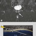
Definitions for synovitis on imaging have been produced by the Outcome Measures in Rheumatoid Arthritis Clinical Trials (OMERACT) initiative, defining synovial hypertrophy on ultrasound as hypoechoic tissue within and involving the joint capsule that is nondisplaceable and poorly compressible and that may exhibit Doppler signal ( Fig. 32.2 ).