Primary malignant tumor of liver composed of primitive mesenchymal cells with partial, divergent differentiation
IMAGING
• Large, encapsulated, spherical mass
• May have peripheral rim of viable, hypervascular tumor
• Often has large complex, cystic spaces with focal hemorrhage
• May show signs of vascular invasion
Hepatic, portal vein, or inferior vena cava (IVC) invasion
• Large subcapsular tumors may rupture
• Nodal or distant metastases are common
Lung and osseous mets are most common
TOP DIFFERENTIAL DIAGNOSES
• Metastases and lymphoma, hepatic
• Hydatid (echinococcal) disease
• Angiosarcoma, liver
CLINICAL ISSUES
• Majority of patients are children (ages 6-10)
• Comprises 6-13% of primary hepatic neoplasms in children
• Rare reports in adults of all ages
• Generally grim prognosis; 5-year survival averages ∼ 15%
• Resection ± adjuvant or neoadjuvant therapy have recently improved prognosis
DIAGNOSTIC CHECKLIST
• Check for primary extrahepatic tumor
• Metastases are much more common cause of complex cystic hepatic tumors
(Left) Axial T2WI MR in a 57-year-old woman shows a huge hepatic mass that replaces most of the right lobe. The mass is heterogeneously hyperintense, though not as bright as another lesion that proved to be a cavernous hemangioma.
(Right) Axial T1WI C+ MR in the same case shows nodular peripheral enhancement of the hemangioma , whereas most of the sarcoma shows no enhancement. The tumor is an undifferentiated sarcoma, which is largely necrotic and hemorrhagic as suggested by MR.
(Left) Axial arterial phase CECT in a 73-year-old man shows a large, encapsulated mass that has central necrosis and a periphery of hypervascular solid tumor, a common feature of sarcomas in general, though not specific for a primary hepatic sarcoma.
(Right) Coronal CT reconstruction in the same case shows the large, centrally necrotic hepatic sarcoma . Absence of immunohistochemical evidence of muscle, epithelial, or vascular differentiation led to the final diagnosis of undifferentiated sarcoma.
TERMINOLOGY
Synonyms
• Undifferentiated embryonal sarcoma
• Malignant mesenchymoma
Definitions
• Primary malignant tumor of liver composed of primitive mesenchymal cells with partial, divergent differentiation
IMAGING
General Features
• Best diagnostic clue
Large, spherical, encapsulated necrotic or complex cystic mass
Only gold members can continue reading. Log In or Register to continue


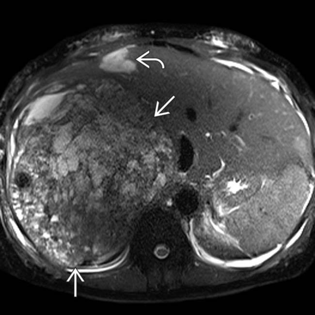
 that replaces most of the right lobe. The mass is heterogeneously hyperintense, though not as bright as another lesion
  that proved to be a cavernous hemangioma.
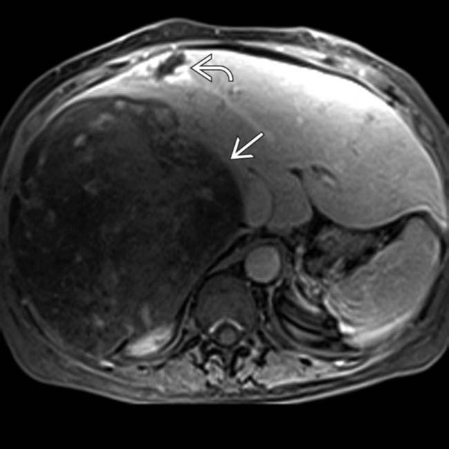
 , whereas most of the sarcoma
  shows no enhancement. The tumor is an undifferentiated sarcoma, which is largely necrotic and hemorrhagic as suggested by MR.
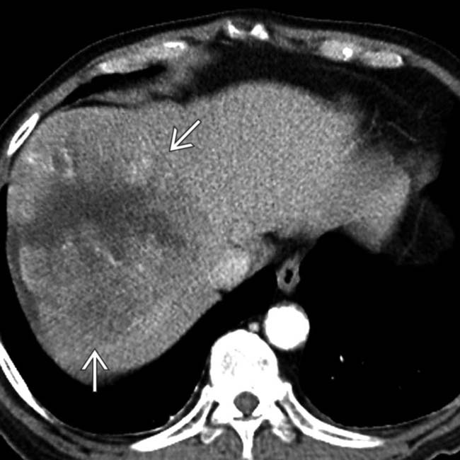
 that has central necrosis and a periphery of hypervascular solid tumor, a common feature of sarcomas in general, though not specific for a primary hepatic sarcoma.
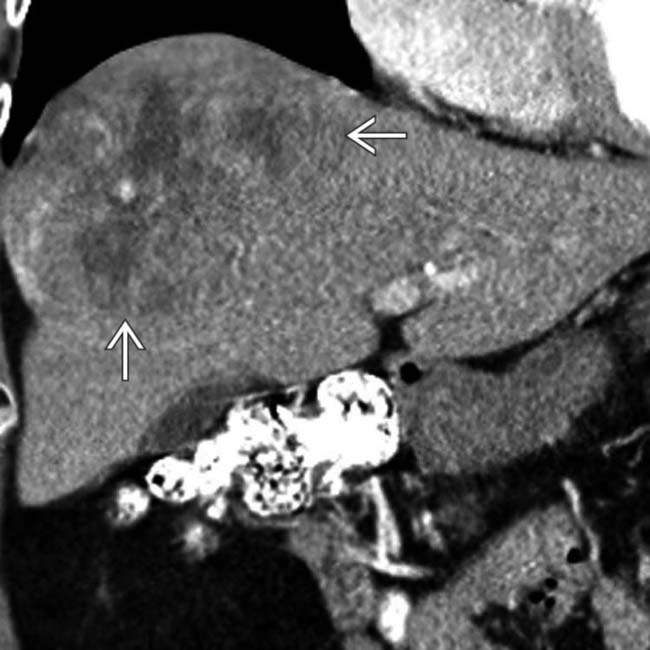
 . Absence of immunohistochemical evidence of muscle, epithelial, or vascular differentiation led to the final diagnosis of undifferentiated sarcoma.


