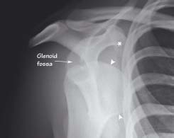
The surface of the humeral head (arrowheads) does not align with the line formed by the anterior rim of the glenoid fossa. There has been no visible bony fracture
22.2 Anterior shoulder dislocation: axial view
The humeral head (arrowheads) is displaced forwards out from the glenoid fossa and now lies under the corocoid process of the scapula (*). This indicates an anterior dislocation
22.3 Pathological fracture
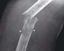
There is a transverse fracture of the mid-shaft of the humerus. There are areas of lucency of the bone trabecular pattern and in places there is loss of cortical thickness (arrowheads). The patient had multiple myeloma
22.4 Clavicle fracture
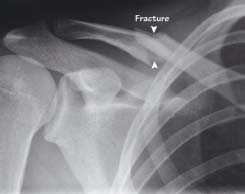
There is a midshaft fracture of the clavicle. The distal part is pulled downwards by the weight of the arm and the proximal part is pulled up by the sternocleidomastoid muscle
22.5 Radial head fracture
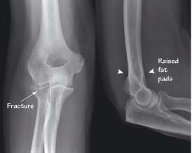
The AP view (left) shows a fracture passing through the radial head into the elbow joint. There are raised fat pads (dark) seen both at the front and back of the distal humerus on the lateral image (right). In the context of trauma it indicates an intra-articular fracture even if no fracture is seen
22.6 Supracondylar fracture
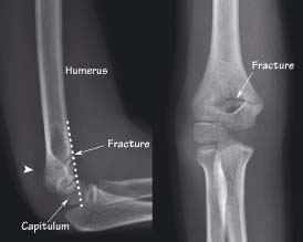
Less than one-third of the capitulum lies in front of the anterior humeral line (dotted), indicating a fracture which is seen clearly on both views. There is a raised posterior fat pad (arrowhead) which is a sign of an accompanying joint effusion
Shoulder dislocation
Anterior
Related posts:


Stay updated, free articles. Join our Telegram channel

Full access? Get Clinical Tree


