A transverse fracture of the distal radius is clearly seen on both views. Dorsal angulation of the distal component and an accompanying fracture of the ulnar styloid (*) are classic features of a Colles’ fracture
23.2 Scaphoid waist fracture: AP view
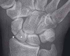
A fracture (arrowhead) passes across the waist of the scaphoid. Failure to treat this injury leads to a high risk of avascular necrosis of the proximal pole (*). This fracture is often not detected on X-ray and so clinical suspicion should lead to treatment with clinical and radiological follow-up
23.3 Monteggia fracture-dislocation: lateral view
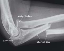
A transverse fracture of the ulna shaft is accompanied by dislocation of the head of radius from the capitulum of the humerus
23.4 Greenstick fracture: AP and lateral views
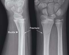
A transverse fracture of the distal radius breaches the dorsal cortex and buckles the ventral cortex. These are typical features of a greenstick fracture
23.5 Boxer’s fracture: AP and oblique views
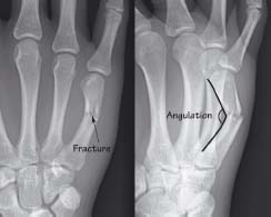
There is a transverse fracture of the little finger metacarpal with palmar angulation of the distal component. This common fracture is said to relate to poor fighting skills. This patient had punched a wall in anger while intoxicated
23.6 Rheumatoid arthritis: both hands
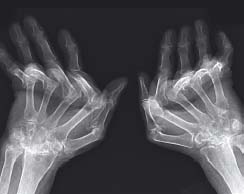
Severe changes of rheumatoid arthritis are shown. These include loss of the carpal joint spaces, erosions of the metacarpal joints and volar subluxation of the metacarpophalangeal joints with ulnar deviation of the phalanges
Distal radius and ulna wrist fractures
- Colles’ fracture
Related posts:

Stay updated, free articles. Join our Telegram channel


Full access? Get Clinical Tree


