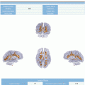
Fig. 16.1
Temporal diagram of the treatment and follow-up of GB patients including (1) the available clinical information at each stage, (2) the treatments (in blue) and (3) the main clinical decisions (in orange). RT dose dist. mean the information about the radiotherapy dose distribution. Q4.n and MR3.n mean the successive decisions and image acquisitions done during the follow-up, respectively
16.3 Main Questions in Glioblastoma Management
Based on the above-mentioned standard clinical workflow, we could identify the following main steps in standard treatment for GB:
Presurgery: to generate a first diagnosis based on medical imaging information
Surgery: to remove the maximum safe area suspected to be affected by the tumour and to analyse the resected tissue to generate a more accurate diagnosis
Concomitant RT with CHTH (based on TMZ): to irradiate the tumour tissue and to avoid the fast propagation of the tumour cells
Follow-up including CHTH as adjuvant treatment to avoid the fast propagation of the tumour cells
In each of these four steps, important clinical decisions have to be addressed in order to select the most adequate treatment for each individual patient. Among them, the key decisions in GB diagnosis, therapy and follow-up are presented in the following subsections:
16.3.1 Presurgery Decision
What is the precise extension of the tumour that determines the maximum area that can be safely resected? Surgery of GB is by definition incomplete given the diffuse infiltrative nature of the tumour and the inability to remove it entirely without causing too much harm to the healthy brain. A major challenge in therapy of GB is the selection of the area for maximum safe resection of the tumour in order to reduce the degree and time to tumour recurrence while at the same time affecting the patient functionality as less as possible [6].
16.3.2 Post-surgery Decision
What is the molecular subtype of GB? What are the implications of GB subtyping in patient prognosis, treatment and follow-up? In recent years, analysis of genomics, transcriptomics and proteomics have identified subtypes of GB with prognostic implications and different responses to treatment. After surgery, it is possible to characterise the molecular subtype of GB using high-throughput arrays and immunohistochemistry techniques. Moreover, liquid biopsy may provide a wide set of biomarkers related to diagnosis, prognosis and treatment response. These biomarkers can circumvent problems of tumour heterogeneity and can be obtained to monitor tumour changes over time. It is now fully clear that different genetic subtypes of GB exist, associated with differences in molecular pathways involved and in biological behaviour. Therefore, clinical questions related to the prognosis and treatment response will be analysed in the context of knowledge of the molecular and genetic underpinnings.
16.3.3 Pre-radiotherapy Decision
What are the best RT dose value, distribution and technique for a specific patient? Currently the RT dose is estimated homogeneously based on anatomical images from PET or magnetic resonance (MR) scanners. The challenge in the use of RT is to reduce the margins beyond the conventional clinical target volume to the minimum in order to have optimised planning target volumes in accordance with the ICRU 62 definitions [7]. A reduction of RT treatment region uncertainty and a better estimation of the RT dose distribution based on the integration of functional information extracted from the images with dose painting could allow reduction of the radiation applied to brain functional areas where necessary and increase of radiation where possible, thereby improving the quality of life of the patients and their survival times.
16.3.4 Follow-Up Decisions
Is the treatment working properly? What should be the duration of adjuvant CHTH? What is the best treatment management during follow-up? Accurate interpretation of MRI scans in terms of the biological evolution of the tumour is an important issue for measuring treatment response both in the setting of clinical trials and in routine clinical care. However, the evaluation of the disease progression still remains a difficult task in the face of treatment modality. Pseudoprogression of tumour versus true progression has become a confusing issue after treatment with TMZ and RT. Radiation injury (radionecrosis) is a potential late complication of RT, especially focal high-dose RT, and can easily be confused with tumour progression. Differentiating the two entities is problematic and often requires long-term follow-up with standard MRI, clinical assessment and use of corticosteroids [1]. By contrast, pseudoresponses may occur after angiogenesis-targeted therapies, as a consequence of changes in vascular permeability. In this sense, an early and accurate assessment of treatment response will improve the decision on maintenance or discontinuation of adjuvant CHTH, as well as the election and timing of subsequent treatments, including second-line CHTH and new local therapies.
16.4 Imaging Biomarkers in Glioblastoma Management
The development of imaging biomarkers is providing new insights into tumour behaviour that were not available from conventional medical imaging. Imaging biomarkers have demonstrated to be relevant for the assessment of tumour grading and response to therapy, without any spatial or temporal constraints. These imaging biomarkers are based on imaging modalities such as PWI, DWI, MRSI and PET.
The inclusion of PWI biomarkers characterising the presence and properties of angiogenesis, vasculogenesis and tumour vascular heterogeneity might improve tumour grading, prognosis and follow-up evaluation [8–10]. The complex modelling of dynamic susceptibility contrast (DSC) MRI sequences has also allowed for the quantification of tumour permeability and angiogenesis processes, through pharmacokinetic models of the lesion.
DWI may allow the cellularity of tumours to be graded noninvasively; because cells constitute a relative barrier to water diffusion, compared with extracerebral space, tumours that are more cellular are expected to show less of an increase in apparent diffusion coefficient (ADC) than tumours that are less cellular [15]. Diffusion tensor imaging and diffusion kurtosis imaging are used to describe diffusion 3D variability by means of mean diffusivity, fractional anisotropy and mean kurtosis. Several studies suggest that diffusion tensor imaging allows not only to observe high cellularity regions but also to evaluate tumour invasion into the surrounding tissue [16]. Studies of patients with brain tumours have shown that increases in water diffusion generally indicate positive response to therapy [15].
MRSI provides information regarding the concentration of specific metabolites throughout the brain, which has proven to be relevant in brain tumour diagnosis and prognosis. Thus, increased lipid levels are found in high-grade gliomas, indicating the presence of necrosis, which is a hallmark of GB [17]. Choline has been related to cell membrane density and is recognised as a marker of cell proliferation [17]. Statistically significant higher metabolite ratios of choline/creatine and choline/NAA have been reported in high-grade gliomas compared to low-grade gliomas [18]. Elevated choline levels have been found in peritumoural oedema surrounding GB, suggesting tumour invasion. After treatment, MRSI has also shown potential to differentiate tumour recurrence from radiation necrosis [19].
Several PET tracers have shown their added value when it comes to the diagnosis, prognosis and treatment monitoring of brain tumours. 18 F-FDG, which is a marker of glucose metabolism, has shown correlation with tumour grade and survival rate in gliomas [20]. Increased amino acid PET tracer uptake has been related to angiogenesis and increased cell metabolism within gliomas, resulting in a higher 11C-MET uptake in high-grade than in low-grade gliomas [21]. Labelled nucleotides such as 18 F-FLT are indicators of cellular proliferation, promoting 18 F-FLT kinetic analyses to assess early treatment response [22]. 18 F-FMISO is a hypoxia marker, showing increased uptake in high-grade but not in low-grade gliomas. Tumour progression and survival after RT have been related to 18 F-FMISO uptake levels [23].
Hypoxia plays a central role in tumour development, angiogenesis, growth and resistance to treatment. Hypoxia measurements have been shown to correlate with the probability of metastatic spread, tumour recurrence, resistance to CHTH and radiation, invasion and decreased patient survival. Only a few imaging techniques have potential for in vivo assessment of hypoxia in humans, particularly for repeated, sequential measurements [24]. These methods use either PET tracers or MRI techniques sensitive to variations in local oxygen changes such as blood oxygenation level-dependent MRI (BOLD-MRI) or dynamic contrast-enhanced MRI (DCE-MRI)). An additional approach to map regional hypoxia is through the use of 3D MRSI and the quantification of lactate to N-acetyl-aspartate ratio with long echo times.
Although the added value of PWI, DWI, MRSI and PET is unmistakable, it has become clear that no single modality in itself is specific enough to show the early response to treatment of GB tumours due to their heterogeneity and evolution speed [4]. Hence, some groups have studied the complementary information provided by different modalities and techniques. Laimon et al. described the complementarity regarding tumour progression and response of dynamic [18 F] fluorothymidine (F-18 FLT) PET, sodium (23Na) MRI and 3-T morphological MRI biomarkers.
Manual segmentation is still the gold standard for brain tumours in clinical practice; however it implies a time-consuming and user-dependent bias, prone to errors and with questionable reproducibility. Significant progresses have been made in automated brain tumour segmentation based on machine learning [25–27]. Brain Tumour Segmentation (BRATS) Challenge on MICCAI Conference revealed that machine learning performs well in the whole tumour segmentation compared to manual segmentation. However, supervised learning requires an expensive, time-consuming and biased task to retrieve a sufficiently large set of labelled samples from which to learn discriminant functions for the posterior segmentation [26]. Moreover, spatio-temporal changes in clinical environment such as new MR machines, protocols or centres may distort the data and hence could affect the performance of the supervised models [28]. Unsupervised learning tackles these limitations in a more straightforward manner, as it directly learns the patient specific data to build an intra-patient segmentation model which is independent from the differences among patients [29].
16.5 New Trends for Integrated GB Management
Current clinical practices in GB management need to evolve to improve the poor results obtained to date in the treatment of this complex disease. To do so, the following promising approaches need to be particularly taken into account.
16.5.1 GB Molecular Subtypes
In the last decade, genomic analyses, transcriptomics and proteomics have identified different GB subtypes and molecular pathways with implications for prognosis and treatment response. For optimal management of patients, more precise classification of gliomas is needed, and molecular markers hold great promises in this respect.
The proneural subtype of GB, which is associated with better prognosis, is characterised by the expression of the histological markers OLIG2, DLL3, PDGFRA, IDH1 mutation (isocitrate dehydrogenase1), the absence of chromosomal gains or losses, the loss of TP53 heterozygosity and the normality of EGFR (epidermal growth factor receptor) as well as PTEN (phosphatase and tension homolog). The mesenchymal subtype, which corresponds to tumours of worse prognosis with strong angiogenic and inflammatory features, is characterised by the expression of mesenchymal markers such as YKL-40, PECAM1 (CD31), VEGF and its receptors one and two, gain of chromosome 7, loss of PTEN, normal or extended EGFR and MET, 17q11.2 deletion as well as high expression of genes of the TNF superfamily and NF-kB (nuclear factor kappa-light-chain-enhancer of activated B cells) signalling pathway. Other less consensual subtypes are proliferative and classic, which share loss of PTEN and frequent EGFR amplification. Proliferative subtype is characterised by histological markers, such as TOP2A and PCNA (proliferative cell nuclear antigen), and loss of chromosome 10. The classic subtype harbours frequent amplification of chromosome 7, loss of chromosome 10, amplification of EGFR gene and absence of alterations in TP53, NF1, PDGFRA or IDH1 [3, 30, 31]. The neural subtype is characterised by neural markers. In retrospective studies, it was observed that classic and mesenchymal tumours benefited from combined treatment of RT plus TMZ, while in the proneural, TMZ did not seem to provide therapeutic benefits [3]. However, prospective studies are necessary to confirm these findings.
On the other hand, recent studies suggest that antiangiogenic therapy could be beneficial in the proneural subtype and possibly in the proliferative subtype, but not in the mesenchymal [32]. All in all, it is currently clear that GB constitutes a ‘mixed bag’ of tumours and that the diagnosis of particular molecular subtypes (especially those characterised by mutations in IDH1/IDH2, H3F3A, BRAF, EGFR variant III (EGFRvIII) or EGFR amplification) will be of relevance in daily clinical practice soon.
Given the complexity and costs of these studies, promising approaches endeavour to develop easy-to-use panels of workable tests in the clinical context able to provide a more precise classification of gliomas, such as the analysis of messenger RNA (mRNA) by Colman and co-workers [8], which identifies nine genes with prognostic value. Another study using immunohistochemistry methods used only three markers for the classification in proneural-like and classical-like subtypes (p53, PDGFRA and EGFR) [33]. In addition, the expression of specific proteins, such as OLIG2, DLL3, TOP2A, CD44, VEGF and FOXG1, has been validated as a feature of GB molecular subtypes. Notably, activation of the signalling pathways pErk1,2/pMAPK and pAKT has also shown its prognostic value in GB [9]. More recently, an innovative, minimal IHC-based scheme for GB subclass assignment was proposed in terms of positive staining for IDH1R132H for proneural, high-EGFR expression for the classical subtype and a combined high expression of PTEN, VIM and/or YKL40 for the mesenchymal subtype [10].
16.5.2 Key Enabling Molecular Biomarkers in the Clinical Practice
Tumour-derived molecular biomarkers include proteins, nucleic acids and tumour-derived extracellular vesicles. These molecular biomarkers are mainly identified in plasma, serum, blood platelets, urine and/or cerebrospinal fluid. These molecular biomarkers provide valuable information of the mechanisms associated with cancer hallmarks such as cell proliferation, tumour progression, invasion, cell cycle, angiogenesis and apoptosis. Recently, circulating tumour cells have also been identified in the blood of glioma patients. Circulating molecules, vesicles, ‘tumour-educated’ platelets and cells may be useful as easily accessible diagnostic, prognostic and/or predictive biomarkers to guide the patient management.
There is an increasing interest in identifying the protein profile of each GB subtype from peripheral blood samples, in addition to the immunohistochemical analysis of a small set of proteins for GB stratification and the activation of key signalling pathways to identify potential therapeutic targets. A different hypothesis suggests that the data obtained will allow the correlation of a simple immunohistological classification pattern with cell-free circulating proteins that may be used to predict prognosis as well as therapeutic response. If this hypothesis is confirmed, it is expected that a wide perspective will be opened for identifying new and more effective therapeutic targets. In this sense, advanced approaches aim to incorporate an accurate selection of molecular biomarkers (e.g. IHC, NGS, methylation status and chromosomal copy number aberrations) including those obtained from liquid biopsy in the clinical management of GB (e.g. cell-free circulating proteins and RNA sequencing of ‘tumour-educated’ blood platelets).
Thereby, these approaches may help to circumvent problems related to tumour heterogeneity and sampling error at the time of diagnosis. If the success of these methodologies is confirmed, it is expected that a wide perspective will be opened for identifying more informative biomarkers with diagnostic, prognostic, predictive and/or monitoring value and innovative, more promising therapeutic targets.
16.5.3 Advanced Multiscale Data Modelling in GB: In Silico Oncology Models
The main approaches for multiscale mathematical modelling of cancer have as a common starting point the fact that cancer is a genetic disease and that its evolution is related, since the very early stage to mutations that give acquired abilities in few or even single cells [34, 35]. The observation that the biological system under consideration has multiscale features has resulted in the development of mathematical models that essentially couple different models operating at different scales and that are able to cope with genomics, proteomics, cell-cell interaction, cell-environment interaction, release, diffusion and absorption of chemical factors. Specifically, the modelling of cancer dynamics at the lowest scale, namely, molecular and cellular scale, focuses on the critical changes within the cell that characterise cancer growth. These changes (i.e. self-sufficiency in growth signals, insensitivity to antigrowth signals, evading apoptosis, limitless replicative potential, sustained angiogenesis, evading immune system attack and tissue invasion and metastasis) incorporate some aspects of genetic mutation, gene expression and evolutionary selection, leading to malignant progression. In various cases, this evolution is induced by external or concomitant actions (as an example, the effect of therapies) [36]. At the tissue scale, macroscopic models of gliomas focus on the heterogeneous and anisotropic characteristics of the brain-deducing models that are able to describe the growth of tumour masses and the diffusion of metastases in such an environment [37, 38].
Related posts:
 The Shift in Paradigm to Precision Medicine in Imaging: International Initiatives for the Promotion of Imaging Biomarkers
 The Final Step: Imaging Biomarkers in Structured Reports
 Introduction to the Stepwise Development of Imaging Biomarkers
 A Proposed Imaging Biomarkers Analysis Platform Architecture for Integration in Clinics
 Use Case III: Imaging Biomarkers in Breast Tumours. Development and Clinical Integration
Stay updated, free articles. Join our Telegram channel

Full access? Get Clinical Tree


