Valvular Heart Disease


  • Cardiovascular magnetic resonance (CMR) can identify valvular lesions, assess their severities, and determine the consequences of the valve lesion on left ventricular function.


  • CMR makes highly accurate and reproducible measurements of ventricular size, mass, and systolic function.


  • CMR can provide additional clinical information in patients who have poor echocardiographic windows.


  • CMR can evaluate for associated vascular abnormalities, such as the thoracic aorta, during the same evaluation.


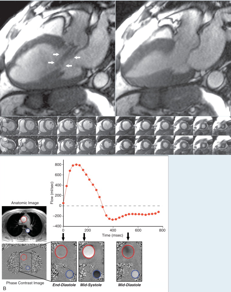
  • Accurate measurements of regurgitant volumes is performed with phase contrast CMR.


  • One strength of CMR is evaluation of myocardial viability or infarction, which may contribute to the severity of valvular disease.


  • Velocity-encoded phase contrast CMR can accurately estimate intracardiac shunts (Qp : Qs).


  • A comprehensive CMR evaluation is not limited by ionizing radiation or nephrotoxic iodinated contrast agents and is well suited for serial assessments to determine optimal time for intervention.


KEY POINTS


Case 1


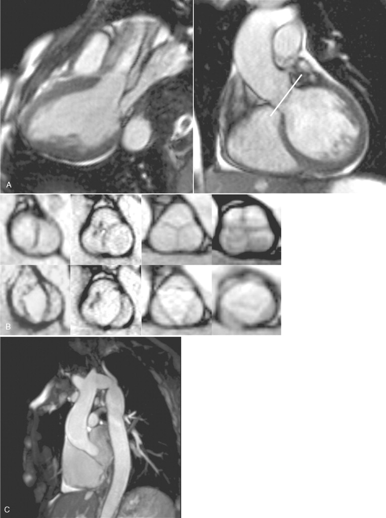
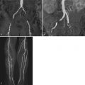
This is a case of a 40-year-old woman with Turner’s syndrome, hypertension, and poor echocardiographic windows ( Figure 7-1 ).


Figure 7-1


A, Systolic three-chamber steady-state free procession cine CMR ( left image ). The bright signal intensity within the ascending aorta during systole is caused by the acceleration of blood through a mildly narrowed aortic valve. Coronal aortic valve localizer image at end-diastole ( right image ). The white line depicts the imaging plane used to prescribe the aortic valve images in a short-axis orientation for panel B. B, Spectrum of aortic valve morphologies with short-axis cine CMR at end-diastole ( top row ) and end-systole ( bottom row ). This patient’s bicuspid aortic valve ( first column ) is compared with functionally bicuspid ( second column ), normal tri-leaflet ( third column ), and quadricuspid ( fourth column ) aortic valves. Note that no raphe is evident at end-diastole in this patient’s true bicuspid valve, whereas the functionally bicuspid valve has a raphe from the fused right and noncoronary cusps. During systole, the shape of the bicuspid aortic valve orifice is elliptical, as opposed to triangularly shaped in a normal trileaflet valve. The quadricuspid aortic valve has a unique X configuration at end-diastole with a square-shaped orifice during systole. C, Thin maximum intensity projection image of the thoracic aorta from a diaphragm-navigated ECG-gated noncontrast MRA demonstrating coarctation of the aorta. The left subclavian artery arises at the level of the coarctation.


Bicuspid Aortic Valve


Comments


Bicuspid aortic valve is the most common congenital cardiac abnormality, occurring in approximately 1% to 2% of the population. Bicuspid aortic valves are frequently associated with diseases of the aorta, including coarctation, dilation, or dissection. Turner’s syndrome is associated with a higher incidence of both congenital bicuspid aortic valves and vascular abnormalities. In functionally bicuspid aortic valves, three commissures are present but one is fused. The fused commissure or raphe can be visualized at end-diastole and the valve appears tricuspid; however, during systole, the orifice is elliptical shaped. Additional clues for a functionally bicuspid aortic valve are an unequal size of the leaflets or eccentric position. In this case example, the truly bicuspid aortic valve has two cusps symmetrically bisecting the aortic root and no raphe at end-diastole. Quadricuspid aortic valves are an uncommon congenital cardiac anomaly found incidentally in approximately 0.013% of echocardiograms. Although quadricuspid aortic valves are a rare finding, they are associated with aortic valve dysfunction.


Most valvular abnormalities visualized by echocardiography can be assessed by CMR. CMR is useful in patients with poor echocardiographic windows. CMR can also provide additional clinical information such as left ventricular volumes, regurgitant fraction, and myocardial fibrosis or viability. High-quality angiographic assessment of the thoracic aorta can also be done without intravenous contrast during the same examination.


Case 2


This is a case involving a 50-year-old male with asymptomatic severe aortic regurgitation ( Figure 7-2 ).


Figure 7-2


A, Cine CMR in a patient with aortic insufficiency. The three-chamber view during mid-diastole show a relatively dark signal intensity in the left ventricular outflow tract ( arrows ) that represents aortic regurgitation. The signal intensity changes are caused by dephasing and loss of signal from rapidly moving blood, but the appearance of regurgitant jets varies widely with different pulse sequences. On SSFP cine CMR, the jet size of aortic regurgitation is frequently less prominent than on color Doppler echocardiography or in-plane velocity encoded MR images. The stack of short axis images can be planimetered to measure left ventricular dimensions, volumes, and ejection fraction. In this case, the left ventricle is severely dilated (end-diastolic dimension 77 mm, end-systolic dimension 57 mm, end-diastolic volume 389 ml) with moderate eccentric hypertrophy (LV mass 283 g or 132 g/m 2 , upper limits of normal less than 91 g/m 2 ). There is mildly reduced global systolic function (LVEF 50%). B, Quantification of ascending aortic blood flow ( red circle ) and descending aorta ( blue circle ) using axial ECG-gated velocity-encoded phase contrast cine CMR. Anatomic and velocity-encoded phase contrast images through the ascending aorta are obtained simultaneously at the level of the main pulmonary artery bifurcation throughout the cardiac cycle. On phase contrast images, a white signal corresponds to cranial flow and a black signal corresponds to caudal flow. The gray signal intensity of the chest wall denotes stationary tissue, and the speckled appearance of the lungs and air outside the chest wall represents noise (regions with too weak a signal for velocity measurements). At end-diastole, there is little flow in either the ascending or descending aorta. At mid-systole, there is the expected cranial flow within the ascending aorta and caudal flow within the descending aorta. However, during mid-diastole, there is aortic flow reversal caused by severe aortic valve regurgitation with caudal flow within the ascending aorta and cranial flow within the descending aorta. By integrating the velocity of all pixels within a region of interest at each time frame, blood flow can be quantified as a function of time with no geometric assumptions. This is possible because CMR can quantify velocity through the imaging plane. The graph displays ascending aortic flow versus time from all 30 velocity-encoded phase contrast images acquired across the cardiac cycle. Positive values ( above the dotted line ) represent forward flow and negative values represent reverse aortic flow. In this patient with severe aortic insufficiency, the net forward aortic volume was 165 ml per cardiac cycle with a reverse volume of 84 ml per cardiac cycle, yielding a regurgitant volume of 81 ml or a regurgitant fraction of 51%.

Only gold members can continue reading. Log In or Register to continue

Stay updated, free articles. Join our Telegram channel

Feb 1, 2019 | Posted by in MAGNETIC RESONANCE IMAGING | Comments Off on Valvular Heart Disease

Full access? Get Clinical Tree

Get Clinical Tree app for offline access