14
Vascular Anatomy above the Diaphragm
 Arteries
The right and left coronary arteries arise off the right and left aortic sinuses. The brachiocephalic artery is the first branch off the arch, which bifurcates into the right subclavian and right carotid arteries. Next, the left carotid artery and, last, the left subclavian artery arise directly off the arch. (See Table 14-1 for variants of normal arch anatomy and Table 14-2 for genetic abnormalities of the thoracic arch.)
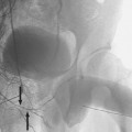
The thoracic aorta is divided into four segments1 (Fig. 14-1): the ascending aorta, the aortic arch, the isthmus (between last great vessel and the attachment of the ductus arteriosus), and the descending aorta. Occasionally, there is a fusiform dilatation of the proximal descending aorta, a ductus diverticulum, which should not be confused with a traumatic injury. Usually, there are nine pairs of intercostal arteries, from the third through the 11th intercostal spaces. There are many variations to the intercostal anatomy. Most commonly, there are between two and four bronchial arteries; multiple bronchial arteries are more common on the left. Bronchial arteries may arise directly off the aorta or have a common origin with an intercostal artery, thus forming an intercostal bronchial trunk.2
The right subclavian artery originates off the brachiocephalicartery and the left subclavian arteryarises directly off the aortic arch. The subclavian artery becomes the axillary artery after it crosses the lateral margin of the first rib. The subclavian artery gives rise to the following: the vertebral artery, the thyrocervical trunk, the internal mammary artery, and the costocervical trunk. There are variants to this anatomy, such as independent origins of branches of the thyrocervical trunk or the costocervical trunk.
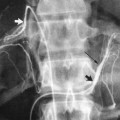
The axillary artery gives rise to several small branches around the shoulder (Fig. 14-2A), including the superior thoracic, thoracoacromial, lateral thoracic, subscapular, and circumflex humoral arteries. There are many variants to this typical anatomy, including absent vessels and independent origins to vessels that may be normally common trunks. Occasionally (2%), the radial artery may originate from the distal axillary artery, and even more rarely (1%), the ulnar artery may originate from the distal axillary artery.
The axillary artery becomes the brachial artery at the lateral margin of the terres major muscle (Fig. 14-2A
Stay updated, free articles. Join our Telegram channel

Full access? Get Clinical Tree


