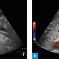
Fig. 1: Position of the probe, subcostal – hepatic veins.
After viewing the portal vein bifurcation one sees the hepatic veins on the lower margin of the image. In terms of depth the image should be focused in a way that one is able to see the diaphragm. It may be necessary to grasp the probe in order to exert sufficient pressure on it. A very clear picture is obtained when the patient inhales deeply.
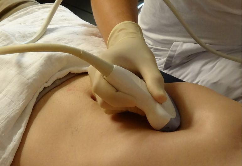
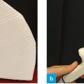
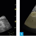
Fig. 2: Position of the probe, subxiphoid – hepatic veins.
If case of difficulty in performing this section, one may use the following trick: move the probe just below the costal archalong the xiphoid. By exerting sufficient pressure one always obtains a very clear view of the hepatic veins in this position.
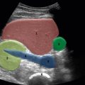
The following structures should be viewed:
- Hepatic veins
- Diaphragm
- Inferior vena cava
Related posts:

Stay updated, free articles. Join our Telegram channel

Full access? Get Clinical Tree


