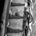
(Left) Prior C7 trauma with extensive anterior cervical fusion and laminectomy. Sagittal T2WI shows instrumented fusion at C4-C5 and fusion at C6-T1 level on a congenital basis. There has been prior laminectomy with large focus of focal cord myelomalacia at C7 seen as cord hyperintensity.
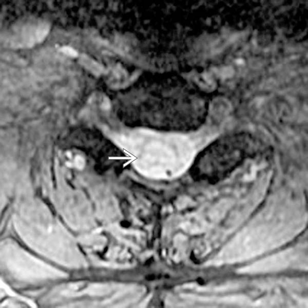
(Right) Axial T2* GRE MR shows the extensive cord myelomalacia at C7 with diffuse increased signal from the cord substance , with essentially no normal cord signal.
(Left) Above the cord lesion, the axial T2* GRE MR shows well-defined high signal intensity wallerian degeneration within the dorsal columns .
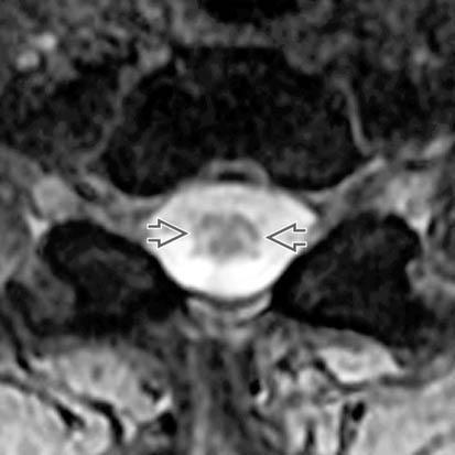
(Right) Below the cord lesion, the axial T2* GRE MR shows high signal intensity wallerian degeneration within the lateral corticospinal tracts .
TERMINOLOGY
Synonyms
• Orthograde degeneration, secondary degeneration
Definitions
• Disintegration of axon and myelin sheath after axonal connection to neuronal cell body is disrupted
IMAGING
General Features
• Best diagnostic clue
Increased T2 signal from dorsal columns above cord lesion; increased signal from lateral corticospinal tracts below cord lesion
• Location
Cervical spinal cord > thoracic
• Size
Variable longitudinal length, generally several vertebral bodies
• Morphology
Linear, following long axis of spinal cord
MR Findings
• T2WI
Increased signal from dorsal columns above site of cord injury
Increased signal from corticospinal tracts below site of cord injury
– Most apparent in cervical cord
Only gold members can continue reading. Log In or Register to continue
 , with essentially no normal cord signal.
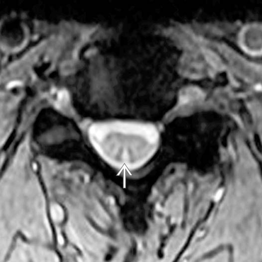
 .
.
 .
.


 Increased signal from corticospinal tracts below site of cord injury