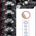
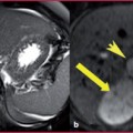
Fig. 14.1
Dynamics of the contrast medium in WB-CTA examinations. After passage into the arterial circulation, the first bolus of the contrast medium (green) is used to scan the coronary circulation (coronary scan), and will come back to the right cardiac chambers, where it will add to the second bolus (in yellow) administered about 40 seconds after the first, becoming available again. So the second bolus will be boosted by the first and enable good opacification of the extra-coronary arterial circle, enabling acquisition of the extra-coronary arteries (extra-coronary scan) by Limiting the total dose of contrast agent used for the examination
Table 14.1
Administration of contrast agents for WB-CTA examinations in the coronary circulation
Coronary acquisition | Extra-coronary acquisition | |
Volume (mL/s) | (Flow rate x acquisition time) + (Speed of flow x break pre-scan) | 40-60 |
Flow (mL/s) | 4-6 | 4-6 |
Saline | From 30 mL/s to 4 mL/s | From 50 mL/s to 4 mL/s |
Delay | Bolus tracking >150 HU | Bolus tracking >150 HU |
The first bolus of the contrast medium, used for coronary acquisition, after its passage in the arterial circulation, returns to the right heart chambers, where it joins the second bolus, ensuring appropriate vascular enhancement of the extra-coronary district.
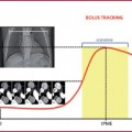
The technical success of the examination is based on an accurate calculation of delay times between administration of the contrast medium and the beginning of the scan. The delay in coronary acquisition is calculated with the bolus tracking technique; after about 40 seconds a relevant fraction of the first contrast agent bolus will be available again for an arterial circulation phase and at that time it will be necessary to administer the second bolus for the extra-coronary examination.
The delay in extra-coronary acquisition is calculated considering circulation parametres: the total delay is, in fact, the sum of the transit time from the venous access to the aorta (the same as for coronary artery acquisition, previously calculated with the bolus tracking technique) and the time of physiological recirculation of the first bolus (approximately 40 s).
If, for example, the delay between the administration of the first bolus and the start of the coronary scan is about 15 s (variable in relation to the patient’s hemodynamic characteristics), the delay for the beginning of the second acquisition will be 15 s + 40 s.
The scanning parameters for coronary acquisition are shown in Table 14.2. The scanning parameters for extra-coronary acquisition are shown in Table 14.3.
Table 14.2
Scanning parameters for the coronary circulation
Coronary circulation | 16 MDCT | 32-40 MDCT | 64 MDCT |
Collimation (mm) | 1.0-1.5 | 0.5-0.625 | 0.5-0.625 |
Slice increment (mm) | 1.0-1.3 | 0.4-0.6 | 0.4-0.6 |
Slice thickness (mm) | 1.5-2.0 | 0.6-0.9 | 0.6-0.9 |
Rotation time | 0.37-0.5 | 0.37-0.4 | 0.33-0.4 |
kV | 120 | 120 | 120 |
mA | 300-540 | 375-540 | 375-600 |
mAs (mA x T rot) | 150-200 | 150-200 | 150-200 |
Effective mAs | 430-800 | 600-800 | 600-990 |
Pitch | 0.25-0.35 | 0.20-0.30 | 0.20-0.30 |
Table speed (mm/s) | 12-16 | 10-15 | 15-22 |
Scan direction | Craniocaudal | Craniocaudal | Craniocaudal |
Table 14.3
Scanning parameters for extra-coronary circulation
Extra-coronary circulation | 16 MDCT | 32-40 MDCT | 64 MDCT |
Collimation (mm) | 1.0-1.5 | 0.5-0.625 | 0.5-0.625 |
Slice increment (mm) | 1.0 | 1.0 | 1.0 |
Slice thickness (mm) | 1.5-2.0 | 1.0-1.3 | 1.0-1.3 |
Rotation time | 0.37-0.5 | 0.37-0.5 | 0.33-0.5 |
kV | 100 | 100 | 100 |
mA | 50-100 |