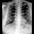
(Left) Thoracolumbar anteroposterior radiograph demonstrates sclerotic focus within the lower thoracic vertebral body, adjacent to the pedicle. There is characteristic nonaggressive appearance with sclerotic density and brush-like margins that suggest bone island.
(Right) Axial bone CT of the thoracic spine reveals a focal sclerotic bone lesion with characteristic dense sclerosis and irregular, brush-like margins of the vertebral body bone island.
(Left) Sagittal T1WI MR demonstrates a small focal area of very low signal intensity within the anterior L4 body , without reactive marrow change or osseous destructive changes to imply an aggressive lesion.
(Right) Sagittal T2WI MR displays a small focal area of very low signal within the anterior L4 body , without reactive marrow change or other worrisome marrow abnormality. Lack of destructive change and soft tissue mass favor bone island.
TERMINOLOGY
Synonyms
• Enostosis, sclerotic island, calcified island, compact island
Definitions
• Asymptomatic focal areas of bony sclerosis
IMAGING
General Features
• Best diagnostic clue
Small focal areas of sclerosis, with feathered or brush-like margins, in cancellous bone
• Location
Found in any skeletal bone
– Most common in pelvis, femur, ribs, long bones
– Less common in spine; may involve vertebral body or posterior elements
• Size
1 mm to several cm
• Morphology
Round, well-defined margins
Radiographic Findings
• Radiography
Single or multiple areas of focal homogeneously dense sclerotic foci in cancellous bone
Distinctive radiating bony streaks (“thorny radiation”) blend with trabeculae of host bone
CT Findings
• NECT
Findings similar to plain radiography, without demonstrable soft tissue component
MR Findings
• T1WI
Focal low signal intensity
• T2WI
Focal low signal intensity
• T2* GRE
Low signal + slight “blooming” effect due to susceptibility artifact
Only gold members can continue reading. Log In or Register to continue
 within the lower thoracic vertebral body, adjacent to the pedicle. There is characteristic nonaggressive appearance with sclerotic density and brush-like margins that suggest bone island.
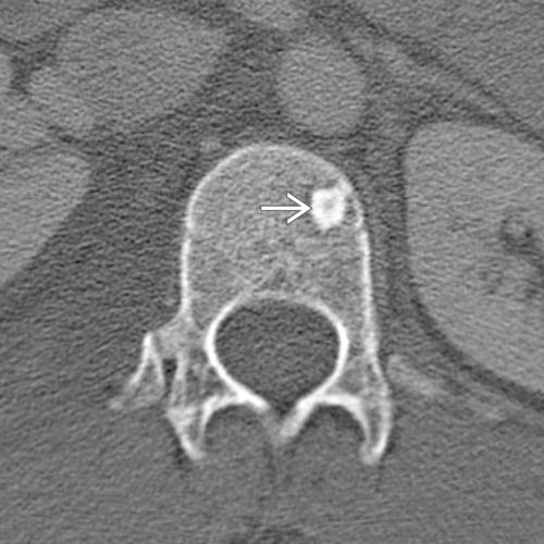
 with characteristic dense sclerosis and irregular, brush-like margins of the vertebral body bone island.
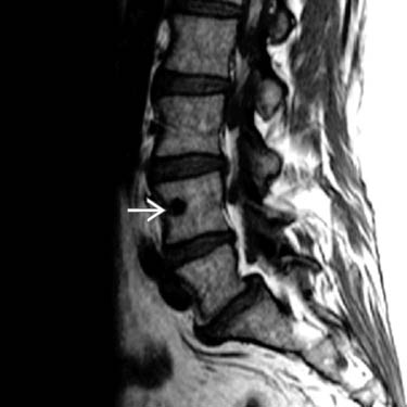
 , without reactive marrow change or osseous destructive changes to imply an aggressive lesion.
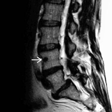
 , without reactive marrow change or other worrisome marrow abnormality. Lack of destructive change and soft tissue mass favor bone island.